

Pancreas
|
Control_Pancreas_HE_03.svs |
Control_Pancreas_Pen_03.svs |
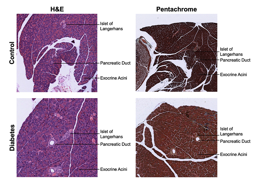
H&E staining demonstrated marked superiority for pancreatic evaluation, providing clear visualization of islets of Langerhans, acinar cells, ductal structures, and nuclear detail. In contrast, Movat pentachrome sections were frequently dark and obscured fine cellular morphology, limiting its utility for diabetes-related assessment despite offering complementary contrast of overall lobular architecture.