
Kidney
 |
 |
|
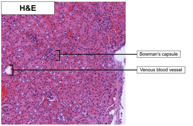
| Kidney_H&E_02.svs Kidney_H&E_02.png |
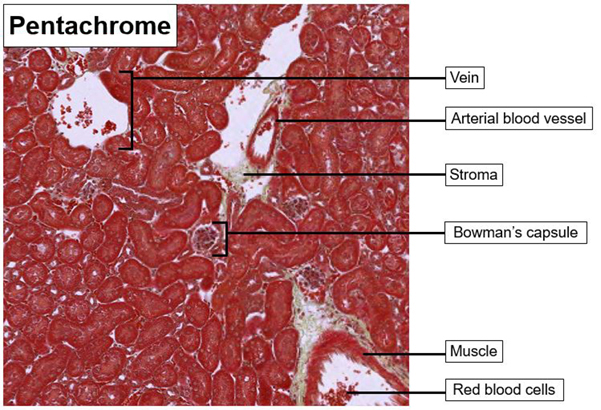
Kidney_Pen_02.svs Kidney_Pen_02.png |
Collagen is more effectively visualized using the pentachrome stain kit at the medulla due to the contrast of the green/blue staining of the supporting stroma. The pentachrome red staining of the muscles helps to differentiate between the type of blood vessels present as the arterial vessels are surrounded by muscle and the venous blood vessels are not.
Previous Attempts
 |
 |
|
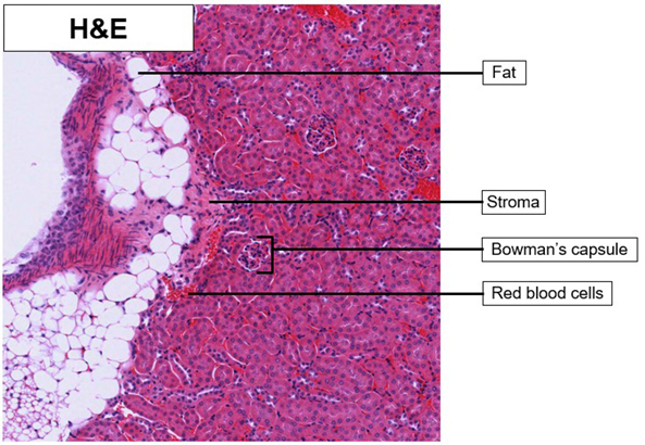
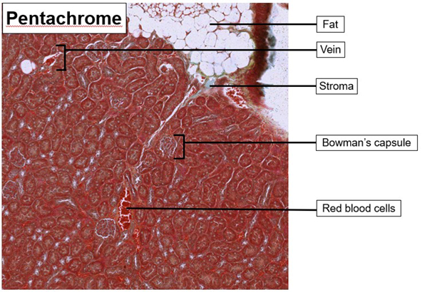
| Kidney_H&E_01.svs Kidney_H&E_01.tif |
Kidney_Pen_01.svs Kidney_Pen_01.tif |
Collagen is more effectively visualized using the pentachrome stain kit at the medulla due to the contrast of the green/blue staining of the supporting stroma. The pentachrome red staining of the muscles helps to differentiate between the type of blood vessels present as the arterial vessels are surrounded by muscle and the venous blood vessels are not. The intensity of the pentachrome red stains on the cytoplasm reduced the visibility of the nuclei. Further, the intensity of the red blood cells stained red obscures the visibility of cellular borders.


