
Heart
 |
 |
|
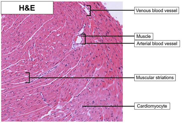
| Heart_H&E_02.svs Heart_H&E_02.png |
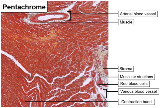
Heart_Pen_02.svs Heart_Pen_02.png |
The pentachrome staining of the stroma (green/blue) is well visualized. The pentachrome red staining of the muscles helps to differentiate between the type of blood vessels present as the arterial vessels are surrounded by muscle and the venous blood vessels are not. The striations of the cardiomyocytes are also well visualized with the pentachrome stain kit.
Please note: contraction bands can look fractured, but the appearance presented is dependent on the state of contraction when the tissue is excised and fixed.
Previous Attempts
 |
 |
|
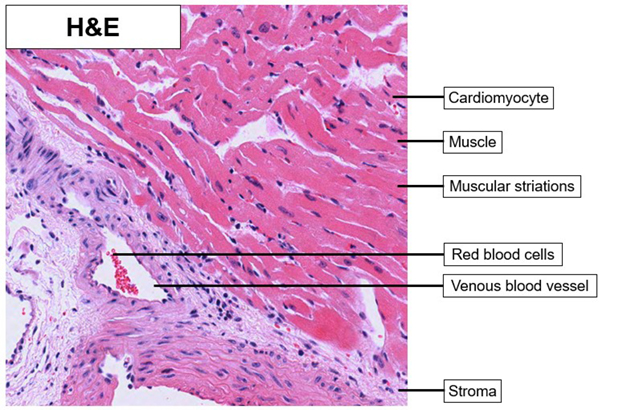
| Heart_H&E_01.svs Heart_H&E_01.tif |
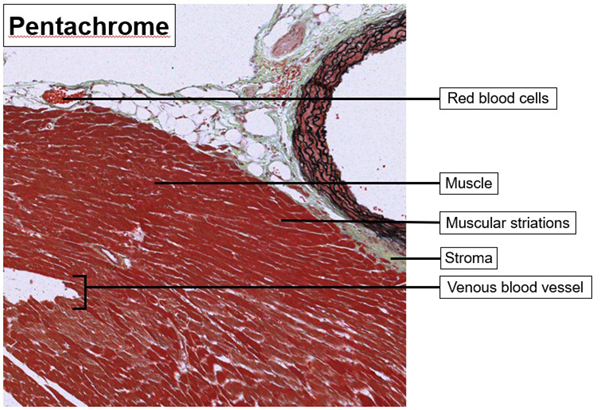
Heart_Pen_01.svs Heart_Pen_01.tif |
Due to the intense pentachrome red stain staining the cytoplasm and muscles, the ability to visualize the cellular structures was obscured. The pentachrome staining of the stroma (green/blue) is well visualized. The pentachrome red staining of the muscles helps to differentiate between the type of blood vessels present as the arterial vessels are surrounded by muscle and the venous blood vessels are not. The striations of the cardiomyocytes are also well visualized with the pentachrome stain kit.
Please note: contraction bands can look fractured, but the appearance presented is dependent on the state of contraction when the tissue is excised and fixed.


