
Eye
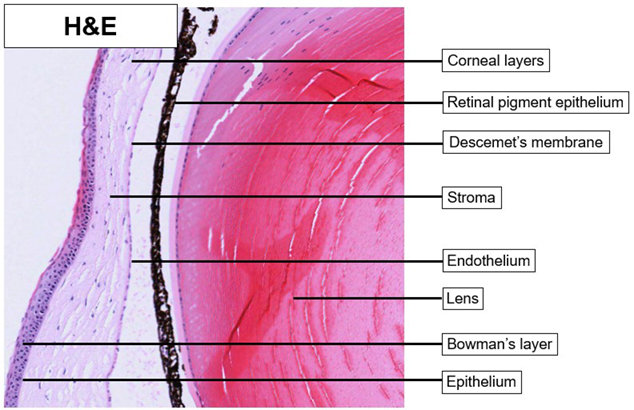
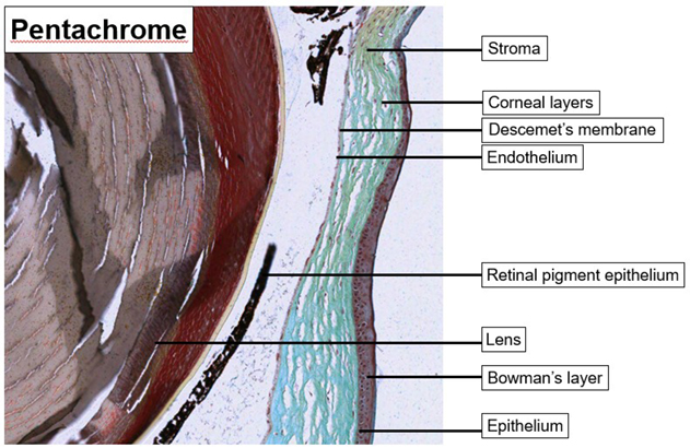
Cornea
 |
 |
|
| Eye_Cornea_H&E_02.svs Eye_Cornea_H&E_02.png |
Eye_Cornea_Pen_02.svs Eye_Cornea_02.png |
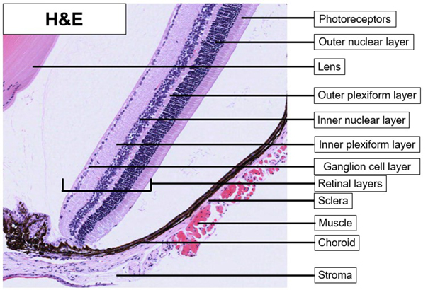
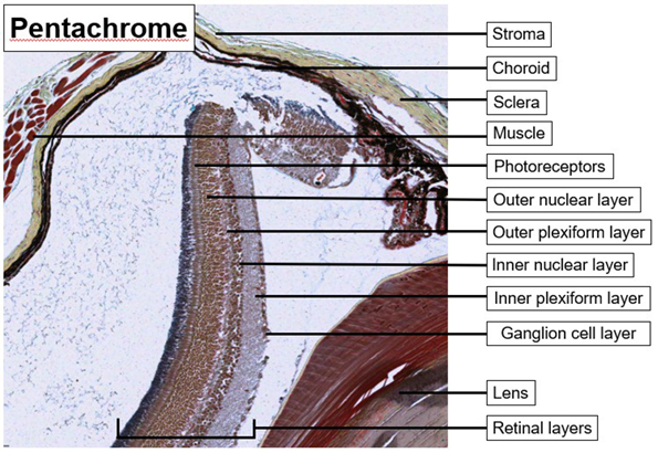
Retina
 |
 |
|
| Eye_Retina_H&E_02.svs Eye_Retina_H&E_02.png |
Eye_Retina__Pen_02.svs Eye_Retina_Pen_02.png |
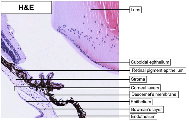
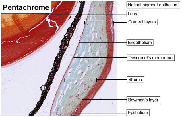
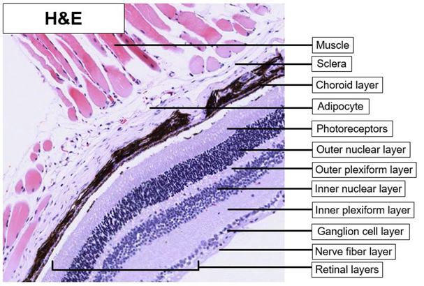
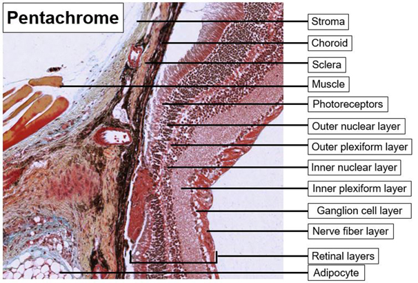
The corneal layers are better visualized using the pentachrome stain kit. The pentachrome stain kit also finely stains the rod cells and allows for easy differentiation of the microstructures of the retina and cornea. The pentachrome stain kit is therefore a finer stain for the fibrillar structures. However, the nuclear staining is not as strong and the inflammatory cells might be less precisely identified.
Previous Attempts
Cornea
 |
 |
|
| Eye_Cornea_H&E_01.svs Eye_Cornea_H&E_01.tif |
Eye_Cornea_Pen_01.svs Eye_Cornea_01.tif |
Retina
 |
 |
|
| Eye_Retina_H&E_01.svs Eye_Retina_H&E_01.tif |
Eye_Retina__Pen_01.svs Eye_Retina_Pen_01.tif |
The corneal layers are better visualized using the pentachrome stain kit. The pentachrome stain kit also finely stains the rod cells and allows for easy differentiation of the microstructures of the retina and cornea. The pentachrome stain kit is therefore a finer stain for the fibrillar structures. However, the nuclear staining is not as strong and the inflammatory cells might be less precisely identified.


