

Adipose
|
Control_Adipose_HE_03.svs |
Control_Adipose_Pen_03.svs |
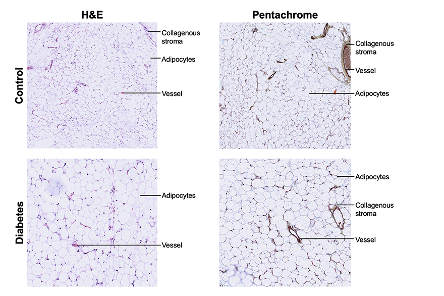
H&E staining provided superior visualization of adipocyte morphology and stromal cell nuclei, facilitating the assessment of cell size and inflammatory components. Conversely, Movat pentachrome more effectively highlighted perivascular collagen, elastin, and vascular walls, offering complementary contrast of the supporting stroma despite less refined nuclear and cellular detail.