

Facilities and Resources
 Intradepartmental facilities
Specialized research facilities available to researchers within Anatomy & Cell Biology include:
Imaging
LiCor Odyssey Classic
Location: MSB 420
An Infrared imaging system. It is an alternative to standard fluorescence imaging given that it is able to eliminate background fluorescence from samples providing clear and sharp bands. Ideal for western blots, this system utilizes direct infrared fluorescence detection to quantify low abundant proteins that other chemiluminescent procedures cannot.Please contact Tom Chrones at tchrones@uwo.ca for details.
Bio Rad Gel Doc XR+
Location: MSB 433
A robust imaging system. Features of the Gel Doc XR+ imaging system with Image Lab image acquisition and analysis software includes:- Quick and accurate gel and blot imaging and analysis
- Automated, hands-off routines that require no training
- The software can save and recall all the steps in the workflow for repeatable and reproducible results
- System optimization at setup, which provides image data that are always accurate, reproducible, and free of imaging artifacts
- A wide range of applications with special accessories that preserve sample integrity for downstream research while ensuring user safety
- Comprehensive, automated, quantitative analysis of protein and DNA samples in seconds
- Reports with customized and organized data
- Quick publication-quality results
- Focused images at all times, regardless of zoom level or sample position
- Appropriate, automatic, and consistent flat fielding correction of image data for every application
- Automatic correction of imaging artifacts
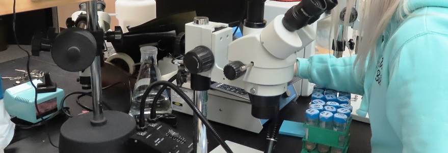
Microscopy
Zeiss AXIO Vert.A1 FL-LED
Location: MSB 418
(transmitted light and LED fluorescence) This microscope is a small and user friendly microscope with phase, brightfield and fluorescence imaging capabilities, using 2 Zeiss Cameras (AxioCam ICc5 and AxioCam 506 mono) and Zen Blue software.Please contact Tom Chrones at tchrones@uwo.ca for details.
Leica Microscopes
Location: MSB 433A
There are 2 Leica microscopes that can be used to image cultured cells (with phase microscopy) and stained slides. The Leica DMIL LED is an inverted microscope, coupled to a Leica DFC295 3MP colour camera, and is well suited for phase an colour imaging. The Leica DM2000 is an upright microscope coupled to a Leica DMC425 5MP colour camera. Both are set up with the Leica Application Suite (LAS) software.Please contact Tom Chrones at tchrones@uwo.ca uwo.ca for details.
Nikon Eclipse Ni-E
Location: MSB 452A
Nikon's top-of-the-line motorized upright microscope, was developed to meet the increasing demands for sophistication and automation of research. Eclipse Ni-E boasts flexible system expandability and supports a broad range of advanced research applications. This microscope is suitable for light and fluorescent microscopy.Please contact Shawn Whitehead at shawn.whitehead@schulich.uwo.ca for details.
Nikon Eclipse Ti
Location: MSB 452A
a very versatile inverted microscope with CrestOptics’ X-Light spinning disk confocal attachment and live cell chamber. This microscope features:- The Nikon Eclipse spinning disk is a high-end confocal live-imaging system suitable for imaging fluorescently labelled cells.
- Heated environmentally controlled enclosure for live imaging applications
- Hardware autofocus prevents stage Z-drift during long time courses
- Extremely sensitive Quantametrics QuantEM:512SC EMCCD camera
- Multi-channel imaging
EVOS M5000 Microscope
Location: MSB 455A
An incredibly easy microscopy solution. The EVOS M5000 microscope is a workhorse for cell imaging labs across a broad range of applications and imaging requirements with minimal requirement for personal contact with the instrument.- No ocular, on-screen display, minimal handling and many more safety features
- No maintenance, assembly, alignment, or calibration
- User defined four-color fluorescence, transmitted light and color applications
- Autofocus, Z-stack capability, time-lapse imaging, live-cell imaging with onstage incubator and single-click multi-channel capture
- On-board software for fast acquisition and analysis, including confluency measurements and automated cell counting
- Access to Thermo Fisher Cloud imaging app
- Semi-automatic, less manual contact
Zeiss AxioPlan 2 Imaging
Location: MSB 419A
Is a upright light/fluorescence microscope. This is unit is operated with the Zeiss Axiovision software (v.4.3). A really nice feature of this microscope is that it can be used manually or with the software to control the objectives and filter sets. This unit is well suited for slide based fluorescence microscopy and phase and DIC imaging.Please contact Tom Chrones at tchrones@uwo.ca for details.
Zeiss AxioVert 40 CFL
Location: MSB 419A
An inverted microscope. This unit is coupled to a Zeiss AxioCam IC1 m camera and the Zeiss Zen Lite software and can be used for phase and fluorescence microscopy with glass slides or TC plates.Please contact Tom Chrones at tchrones@uwo.ca for details.
Molecular Biology
BioRad CFX Connect CFL
Location: MSB 420
A 96-well QPCR-instrument. The CFX Connect Real-Time PCR Detection System offers two-target analysis, excellent thermal cycler specifications, reliable performance and easy set up. For more information please see: https://www.bio-rad.com/webroot/web/pdf/lsr/literature/Bulletin_6105.pdfPlease contact Tom Chrones at tchrones@uwo.ca for details.
Epoch by BioTek
Location: MSB 420
A BioTek microplate reader. With its monochromator-based optics, the Epoch™ Microplate Spectrophotometer offers a filter-free, wide wavelength range for UV-Vis absorbance measurements in a variety of microplate formats, and in 2 µL samples when the available Take3 plate is used. Epoch is controlled with the Gen5 Software interface, with simple programming and powerful data analysis. This robust, low maintenance microplate spectrophotometer is the most cost-effective system available, ensuring even greater value over time.Please contact Tom Chrones at tchrones@uwo.ca for details.
Sample Preparation and Analysis
Singer Instruments ROTOR HDA
Location: MSB 437
This is a Rotor Arraying Robot. It can be used to duplicate and back up large libraries of yeast, fungi, bacteria and algae. It can replicate, mate, and re-array from single or multiple source plates at densities of 96, 384, 1536 and 6144.To use it or to get more details, please contact Patrick Lajoie at plajoie3@uwo.ca for details.
BD FACS Celesta flow cytometer
Location: MSB 418
Which is equipped with Violet (405 nm), Blue (488 nm) and Yellow-Green (561nm) lasers. It also has a high content sampler to allow user to sample specimens from 96 and 384 well plates.To use it or to get more details, please contact Patrick Lajoie at plajoie3@uwo.ca for details.
CryoStar NX-50
Location: MSB 452
This is available to users for routine cryosectioning.Please contact Shawn Whitehead at shawn.whitehead@schulich.uwo.ca for details.Code Repository
Interdepartmental Facilities
Core research facilities available to researchers within the Schulich School of Medicine & Dentistry include:
BioCORE
BioCORE offers services in three well established platforms with high versatility, which serve as tools for custom-designed solutions. Protein Production & Synthetic Biology, Structure, Function & Interaction and High-Thoughput Molecular Analysis.
- For more information visit the BioCORE website
Brain Tumour Tissue Bank
Supplies brain tumour tissues to researchers across Canada and all over the world.
- For more information visit the Brain Tumour Foundation Website
Centre for Functional and Metabolic Mapping
The Centre for Functional and Metabolic Mapping utilizes state-of-the-art 3T whole-body, 7T head and 31 cm 9.4T horizontal MR imaging systems to investigate in-vivo studies of brain structure and function.
- For more information visit the CFMM Website
CSTAR (Canadian Surgical Technologies and Advanced Robotics)
CSTAR provides access to world-class acute and chronic laboratory facilities that support preclinical testing and validation of emergent medical device technologies.
- For more information visit the CSTAR Website
Nordal Cyclotron & PET Radiochemistry Facility
The equipment in the facility includes:
- GE PETtrace 8 cyclotron with proton and deuteron acceleration capability
- cGMP specification class 100 shielded hot cells for production and dispensing
- Dedicated automated chemistry units for producing 18F and 11C radiopharmaceuticals to cGMP specifications
- Availability of 13N ammonia and 15O tracers
- cGMP validated quality control instrumentation
- Synthetic organic chemistry laboratory space
In combination with the PET/CT, PET/MRI, preclinical PET imaging scanners, the facility will be available to support a wide variety of city wide research projects including PET imaging applied to oncology, cardiology, neurology, psychology, bioelectromagnetics, and other areas.
- For more information visit the Lawson Health Research Institute Website
Experimental Transplant Pathology Laboratory
Laboratory provides standardized routine histology, electron microscopy, immunohistochemistry for various species such as monkey, baboon, pig, dog and rodents at the light microscopic or at the ultra structural level, in situ hybridization, apoptosis and morphometric analysis. This facility also provides interpretation with regards to histopathological changes.
- For more information visit the Laboratory website
Fluorescent Deconvolutional Microscope
Our Olympus IX81 fluorescent microscope system with a Coolsnap HQ2 photometrics camera is capable of acquiring multiple fluorescent and white light (DIC) images simultaneously.
- For more information visit the Roth|McFarlane Hand & Upper Limb Centre Website
ICES Western
ICES Western is a satellite site of the Institute for Clinical Evaluative Sciences (ICES) located in Toronto. ICES is an independent, non-profit organization, whose core business is to conduct research that contributes to the effectiveness, quality, equity and efficiency of health care and health services in Ontario. With the establishment of ICES Western, investigators and trainees who wish to pursue research aligning with the ICES mission now have the opportunity to access the large administrative data holdings held by ICES.
- For more information visit the ICES Western Website
Imaging Facilities at the Lawson Health Research Institute
Imaging facilities at Lawson include:
- PET/MR Imaging
- 2 Tesla MRI/MRS
- 3 Tesla MRI/MRS
- Bioelectromagnetics Facilities
- Optical Imaging
- Nuclear Medicine
- 1.5 Tesla MRI
- For more information visit the Lawson Health Research Institute Website
Imaging Research Laboratories at the Robarts Research Institute
The Imaging Research Laboratories at Robarts focus on the discovery and development of innovative imaging techniques and instrumentation to improve the understanding, diagnosis and treatment of human diseases including cancer, cardiovascular and orthopedic disease, schizophrenia and other brain disorders. Using x-ray, ultrasound, CT and MRI, the group is the largest in the Institute, conducting research across nine broad themes:
- basic imaging science & engineering
- brain and mind imaging & spectroscopy
- cancer imaging
- cardiovascular imaging
- Clinical Imaging Research Laboratories
- image-guided surgery & therapy
- molecular, cellular and micro-imaging
- musculoskeletal imaging
- respiratory imaging
- For more information visit the Robarts Research Website
Lawson Clinical Research Services
A versatile, fully-staffed clinical trials facility established by the Lawson Health Research Institute provides increased clinical trials capacity to investigators from London Health Sciences Centre, St. Joseph's Health Care London and community physicians and dentists.
- For more information visit Lawson Clinical Research Services
LHSC Sleep Laboratory and Sleep Clinic
The laboratory and clinic focus on the diagnosis and treatment of adults with sleep disorders, and on clinical research. State of the art, computerized polysomnographic hardware and software are used for the diagnosis of sleep disorders.
- For more information visit the Sleep Lab Website
London Regional Genomics Centre
The facility provides state-of-the-art services on a fee-for-service/cost recovery basis and offers Affymetrix GeneChip technologies and Next Generation Sequencing on the Ion Torrent PGM and Illumina MiSeq platforms. Data analysis software, training and services are also available.
- For more information visit the LRGC website
London Regional Microscopy Facility
The London Regional Microscopy Facility (LRMF), at Robarts Research Institute, Schulich of Medicine & Dentistry, Western University, houses many cutting-edge multiphoton and laser-scanning confocal microscopes; providing paramount systems for both fixed tissue and intravital imaging. The LRMF is dedicated to providing affordable, high quality, accessible, and user-friendly microscopy services for users in Southwestern Ontario. The facility is available to all researchers at Robarts and Western, in addition to members of the Southwestern Ontario research community.
The LRMF's flexible experimental approaches and techniques enable researchers to meet the diverse molecular imaging needs each unique project. Our staff provides complete training and assistance to all users.
- For more information visit the Facility website
London Regional Flow Cytometry Facility
The LR-FCF provides technical and physical support for scientists wishing to utilize flow cytometry analysis and or cell sorting as part of their research. Users are able to utilize the LR-FCF for a wide variety of flow cytometry techniques, including cell sorting, immunophenotyping, cell cycle analysis, cell proliferation, gene transfer (efficacy and tracking), cell death (i.e. apoptosis), cell kinetics, activation states, redox states, chromosome analysis, and cytokine detection among others.
- For more information visit the London Regional Flow Cytometry Facility website
London Regional Transgenic and Gene Targeting Facility
The TGT Facility provides services including:purification of DNA fragments for microinjection; generation of founder transgenic mice; gene targeting services; embryo cryopreservation and rederivation by embryo transfer.
- For more information visit the Facility website
London Tumour Biobank
The London Tumour biobank is a tissue biorepository housed and maintained at the Lawson Health Research Institute. Its goal is to bank snap-frozen biological samples beginning at the time of initial diagnosis, and provide initial diagnostic, therapeutic, and follow-up clinical data to facilitate cancer research across London.
Molecular Pathology core facility
The Molecular Pathology Core Facility entails a full spectrum of state-of-art histopathology equipment and expertise. The facility enables investigators to study fresh, frozen, or paraffin-processed tissues and analyze sections using various histological stains and molecular probes. In addition, micro-isolation of discrete cells within tissue sections can be accomplished using laser capture microscopy.
- For more information visit the Molecular Pathology website
Near Infra-red Spectroscopy
Cerebral blood flow measurement in small animals and neonates.
- For more information visit the Near Infra-red Spectroscopy website
Ontario Tumour Bank
London Health Sciences Centre is one of four sites for the Ontario Tumour Bank, a province-wide biorepository and data bank focused on collection of tumour-related human biospecimens. It provides academic and industry cancer researchers with a diverse selection of high quality tumour-related specimens and data obtained directly by dedicated tumour bank staff, who follow a stringent set of procedures and ethical guidelines.
Pathology and Laboratory Medicine
Pathology and Laboratory Medicine provides a comprehensive range of routine and specialized laboratory testing and clinical consultation to support diagnosis and monitor treatment of patients within London, Southwestern Ontario, nationally and internationally. In addition, Pathology and Laboratory Medicine provides educational training opportunities and continuing education for a broad range of health care professionals. The laboratories also support clinical and applied research carried out in LHSC, SJHC, London Regional Cancer Program, Robarts Research Institute, and Siebens-Drake Medical Research Institute
- For more information visit the Pathology and Laboratory Medicine website
Screening Lab for Immune Disorders
Services include screening tests to evaluate cellular responses in various clinical settings, including autoimmune diseases, inflammatory disorders/conditions, and drug reactions.
- For more information visit the Screening Lab for Immune Disorders
Translational Imaging Research Facility
The 3T MRI Core Facility is a unique regional resource for state-of-the-art evaluation using a variety of MRI techniques in a research setting. The Facility is used primarily for in-vivo studies of human and animal structure and function in the areas of respiratory, cardiovascular, orthopedic and neuro. The HeliSpin™ core facility processes helium-3, an investigational tracer gas used for magnetic resonance (MR) imaging of the lungs which is not possible with conventional (proton) MR imaging.
- For more information visit the Robarts Research Website
Virtual Augmentation and Simulation for Surgery and Therapy (VASST lab)
This facility comprises a high performance computing facility optimized for simulation and visualization of procedures involved in image-guided surgery and therapy. It houses a range of visualization, haptic and tracking systems as well as 2D and 3D
ultrasound equipment.
- For more information visit the VASST Lab website


