

Researchers reveal link between Alzheimer's disease and sex hormones
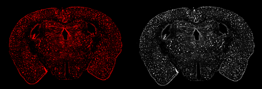 Brain plaques of mouse models in Alzheimer's disease show beta-amyloid protein buildup in the form of bright spots across the brain. (researcher supplied photo)
By Prabhjot Sohal
Alzheimer’s disease disproportionately affects women, who represent about two-thirds of those diagnosed with the late-onset type of the disease.
Previous research has shown Alzheimer’s is also more severe and progresses more rapidly in women, and women with Alzheimer’s experience a steeper cognitive decline – loss of memory, attention, and the ability to communicate and make decisions – compared to men with the disease.
The biological bases for these differences between men and women with Alzheimer’s disease are not well understood. However, understanding them is necessary for developing appropriate therapies.
In a new study in mice and humans, Western researchers have shown female sex hormones play a significant role in how Alzheimer’s manifests in the brain.
The study, published in Alzheimer’s & Dementia: The Journal of the Alzheimer’s Association, also highlights the importance of developing therapeutic strategies focused on these hormonal connections. The research indicates a need to better understand the role of estradiol – a form of the female sex hormone estrogen, used therapeutically to mitigate menopause symptoms – in Alzheimer’s disease.
While the significance of the findings is paramount, the methodology behind them is equally critical, pointing to a necessary shift in scientific approaches.
“To understand how sex hormones play a role in Alzheimer’s, we need to study appropriate animal models. Unfortunately, most studies at this level still focus mainly on the male brain. Our research emphasizes the importance of using animal models that reflect, for instance, postmenopausal women, to understand how sex hormones influence Alzheimer’s pathology,” said Vania Prado, professor, departments of physiology and pharmacology and anatomy & cell biology at Schulich School of Medicine & Dentistry and scientist at Robarts Research Institute.
This study was led by graduate student Liliana German-Castelan, under the supervision of Vania Prado.
Alzheimer’s and the communication system of the brainOne of the key markers of Alzheimer’s disease is the toxic build-up of the protein beta-amyloid in the brain, which eventually disrupts the brain’s communications system and impacts cognition.
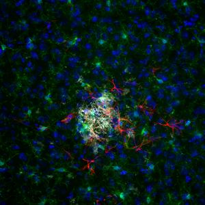 Close-up image of beta-amyloid aggregate in a brain with Alzheimer’s disease. (supplied photo)
The new study shows that the brain chemistry of male and female mice regulates beta-amyloid protein in Alzheimer’s in different ways, with the hormone estradiol contributing to this variation.
Previous studies on mice and at-risk older individuals have revealed that cholinergic neurons, a type of brain cells that produce the chemical messenger acetylcholine, are particularly vulnerable to the damaging Alzheimer’s-associated beta-amyloid accumulation in the brain. Additionally, acetylcholine has been shown to be essential for normal memory and cognition.
While beta-amyloid aggregation impacts the production of acetylcholine, the subsequent loss of this chemical messenger further increases Alzheimer’s pathology, creating a vicious loop.
The team of Western researchers studied this interaction between changes in brain chemistry and the beta-amyloid protein build-up seen in brains impacted by Alzheimer’s.
“Since male and female brains have differences in the cholinergic system, we wanted to see if sex affects this relationship between acetylcholine signalling and the beta-amyloid protein buildup,” said Marco Prado, professor, departments of physiology and pharmacology and anatomy and cell biology. Marco Prado, one of the authors of the study, is also the Canada Research Chair in Neurochemistry of Dementia and a scientist at Robarts Research Institute.
From bench to real world: Representation of sexes mattersIn this study, the researchers observed differences in beta-amyloid accumulation in male and female mice when changing the levels of cholinergic activity. Additionally, they analyzed brain MRI images of healthy older humans.
Different from most studies in humans, in which the MRI scans of man and women are analyzed together, Western professor Taylor Schmitz and graduate student Hayley Shanks analyzed MRI brain scans and the rate of brain loss for aged men and women independently.
 (From left) Graduate student Liliana German-Castelan, and professors Marco and Vania Prado. (supplied photo)
“We observed that the relationship between the integrity of the brain region where cholinergic neurons reside and beta-amyloid accumulation was the same for men and women but was different in male and female mice,” said Marco Prado. The researchers suspected that the fact the female mice being studied were not post-menopausal, while women were, could be an attributing factor to the difference.
The lead author of the study, German-Castelan, intrigued by the sex differences, decided to introduce another layer of testing into the mouse models and with the help of Western professor Robert Gros studied female mice who were closely modelled to represent postmenopausal women. This was done to investigate how the presence or lack of sex hormones could impact the relationship between cholinergic signalling and the beta-amyloid buildup in the brain.
“We found that when the sex hormone estradiol was present, the relationship between acetylcholine and toxic amyloid was lost, but when sex hormones were eliminated in the female mice that relationship reproduced the results seen in humans,” said German-Castelan.
These findings also point to the urgent need to study amyloid and cholinergic function in the ‘peri-menopausal’ age range of 40-50 years, which is much younger than the individuals examined in most large-scale studies of Alzheimer’s disease. Indeed, the sample examined in this study were closer to the age of 70 on average.
“Which explains why there were differences between the results of male and female mice and men and women in our initial exploration,” said German-Castelan.
Researchers emphasized that if they hadn’t included female mice in the study, they might have missed crucial information about Alzheimer’s and sex differences.
“Women and men respond differently to medications and have a somewhat different journey in Alzheimer’s. To develop more effective therapeutics, we need to study animal models that can reproduce different aspects of the journey. Sex hormones and estradiol levels are just one of these factors,” said Vania Prado.
Other authors on the study include Western researchers Lisa M. Saksida and Timothy J. Bussey, and Takashi Saito and Takaomi C. Saido of RIKEN Center for Brain Science, Japan.
Click here for the full manuscript.


