

New 3D ultrasound may improve accuracy of liver cancer treatment
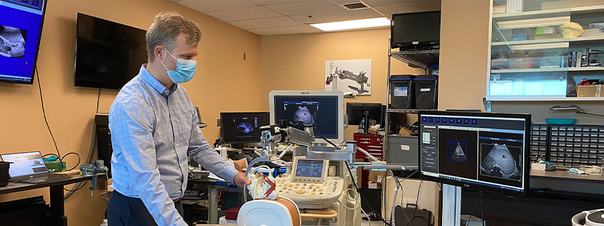
Dr. Derek Cool demonstrating the new robotic 3D ultrasound system. (Lawson Health Research Institute)
By Lawson Health Research Institute
A simulated study by researchers at Schulich School of Medicine & Dentistry and Lawson Health Research Institute has found a new system that uses ultrasound to construct 3D images that could make treatment of liver cancer, using thermal ablation, more accurate.
Liver cancer is the fourth leading cause of cancer death globally. While surgery is one treatment option, thermal ablation – using heat to destroy the cancerous tumour – can have fewer complications and a shorter recovery time. It can also be used for patients who are not surgical candidates. Thermal ablation requires precise needle placement to treat the cancer without damaging the vital organs and blood vessels around it.
“It's very important that we get the needle right in the centre of the tumour,” said Dr. Derek Cool, assistant professor at Schulich Medicine & Dentistry, associate scientist at Lawson, and interventional radiologist at London Health Sciences Centre (LHSC). “If the treatment area doesn't fully cover the tumour, patients are left with a small amount of residual cancer, risking recurrence and the need for additional treatment.”
Ultrasound or CT (computerized tomography) imaging is normally used to guide needle placement, but both are limited. Ultrasound is widely available and can be done in real-time, but only delivers a 2D image. While a CT scan provides a 3D image, it isn’t in real-time and can be a lengthy process.
“We developed a new 3D ultrasound method that shows promise in analyzing whether the complete liver tumour will be ablated by the procedure,” said Dr. Aaron Fenster, professor at Schulich Medicine and scientist at Robarts Research Institute. “And we're now using the same system to guide the needle directly into the centre of the tumor.”
To create the 3D ultrasound images, a robotic cradle moves a standard ultrasound probe, collecting images and stacking them like puzzle pieces.
The simulated study, published in IEEE Transactions on Medical Imaging, used data from 14 patient cases at LHSC to analyze accuracy of the technology. It found that with standard imaging, 64.3 per cent of cases showed complete tumour ablation, while the new system could result in complete coverage for 92.9 per cent of cases (13 of 14 cases). The researchers found that the remaining case could benefit from increasing the ablation time or intensity.
“Our next step is to move from simulation studies to a clinical trial,” said Cool.
If proven effective, the robotic ultrasound system’s portability could potentially allow for more widespread use of 3D ultrasound imaging, including in smaller health care centres. By eliminating the need for CT scans, it could also help to reduce imaging wait times.
“If a clinical trial shows the approach is more accurate and more precise than conventional techniques, there would be a direct impact on patient care,” said Fenster. “We hope to explore commercialization to license the technology and distribute it worldwide.”
 Aaron Fenster, PhD, professor at Schulich Medicine & Dentistry and scientist at Robarts Research Institute
 Dr. Derek Cool, professor at Schulich Medicine & Dentistry


