

Feature: State-of-the-art equipment to advance translational research
A major donation of imaging equipment from industry partners Canon Medical and Carestream is set to advance innovative translational research.
Now installed at Robarts Research Institute, the equipment exactly mirrors what is found in the region’s clinical settings and will support collaborative, multidisciplinary research that bridges from bench to bedside.
The resulting research could improve the outcomes of patients with neurological, cardiac, pulmonary, oncological and musculoskeletal diseases.
“What we’re doing here is fairly unique in Canada,” said Dr. Narinder Paul, Chair/Chief, Department of Medical Imaging. “The projects we will undertake here are multidisciplinary. Imaging scientists can bring their ideas to a project, physicians can address their clinical needs and bring their experience of the current gaps in health care provision, and industry partners can collaborate to move their technologies forward.”
These synergies align with the School’s new five-year strategic plan and illustrate a number of goals focused on new infrastructure and new partnerships.
CT Scanner
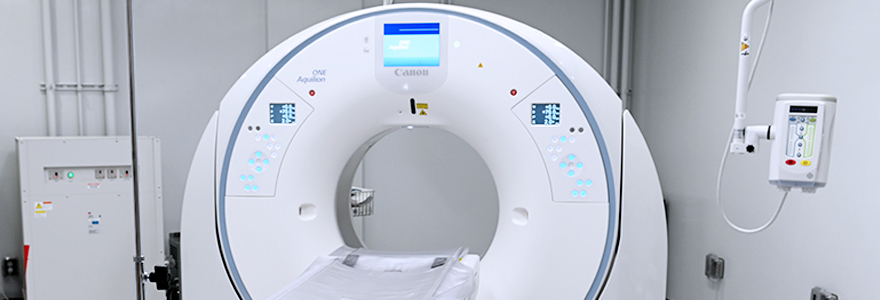
 The Canon Aquilion ONE PRISM Edition CT system in the new centre is the exact same one found in clinical departments at all three hospital sites in London. It will allow researchers to move discoveries more rapidly into clinical practice.
“This is really cutting-edge equipment,” said Matthew Teeter, PhD, Associate Professor, Medical Biophysics. “The Canon CT system is the best in the world right now, and we have it. Combined with fantastic imaging investigators, having the best tools at our disposal should really increase our research impact even further.”
Angiography System
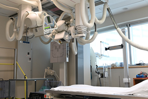
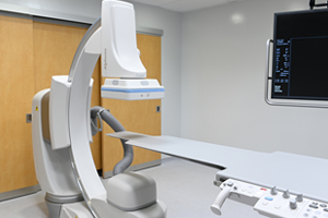 The Canon Alphenix Core+ digital angiography system allows researchers to take video x-rays, including the head-to-toe and fingertip-to-fingertip coverage required to perform various interventional procedures.
It’s the same model often found in hospitals worldwide and used for many interventional radiology procedures, such as inserting cardiac catheters.
The equipment can also become a CT scanner that can rotate around the subject, allowing researchers to render 3D images.
“The Canon angiography system is found across the world,” Paul explained. “Whatever important research we do there, it can be immediately translated into clinical practice.”
Weight-Bearing CT Scanner
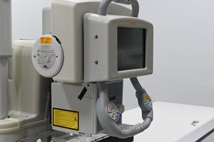
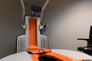 The Carestream OnSight 3D Extremity Scanner is equipped with three x-ray sources to deliver high resolution images.
Unique to the scanner, it allows patients to be scanned in a variety of weight-bearing positions to assess the lower extremities under a natural load.
“Often people or patients will complain of certain pain only when they weight bear. This machine gets the patient standing, and then we can do high-resolution CT scans, so it’s very useful,” Paul explained.
Radiostereometric Analysis Laboratory
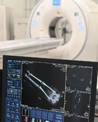
 The RSA Lab features x-ray equipment that allows researchers to assess micromotion in orthopaedic implants for up to two years post-surgery.
“This equipment provides really powerful, quantitative imaging so we can say to a high degree of specificity what is going on inside patients,” Teeter said. “There’s only a handful of centres around the world that can do this type of imaging.”
Digital X-Ray, Dual Energy and Digital Tomosynthesis Suite
 The Carestream DRX-Evolution incorporates high resolution digital X-Ray, state of the art Dual Energy acquisition and Digital Tomosynthesis image acquisition.
“The combination of these features facilitates comprehensive high-resolution structural and functional imaging, combined with tomographic capabilities that improve on conventional X-Ray images by removing overlapping structures and, in certain scenarios, provides information similar to CT but at a much lower radiation dose” said Teeter.
Streamlining and improving patient care
Another major benefit of the new centre is combining multiple imaging modalities under one roof. Teeter said this will directly benefit patients and those involved in studies as they won’t have to travel to multiple sites across the city to be assessed using different equipment.
“You can get so much more information in one visit, so time is maximized,” he said.
The centre also furthers collaborations between scientists, clinicians and technology developers and allows for imaging equipment to be used in novel ways, leading to greater improvements in patient care.
“As researchers, it allows us to dream in a certain way,” Teeter said. “We have all these modalities, and now we can think about which will give us the best information and how we can combine them together to give a picture many wouldn’t have.”
“It all comes back to why we are doing research,” Paul said. “Everything, we do is focused on making life better for patients.”


