

New target identified for preventing bone destruction
Wednesday, December 4, 2013
The skeleton is constantly being remodelled by the breakdown of old bone by cells called osteoclasts and the formation of new bone by cells called osteoblasts. This coordinated activity is essential for maintaining healthy bone. However, excessive osteoclast activity leads to bone destruction in skeletal diseases such as osteoporosis, rheumatoid arthritis and cancer metastases in bone. A family of signaling enzymes known as phosphatidylinositol 3-kinases (PI3Ks) control diverse cell functions but, up until now, little was known about the function of specific PI3K isoforms in osteoclasts.
A paper published in the December 6 issue of The Journal of Biological Chemistry announces the characterization of a new potent and selective PI3Kdelta inhibitor, GS-9820. The discovery was made by members of Western University's Bone and Joint Initiative with collaborators from Nihon University, the University of Calgary and Gilead Sciences, Inc.
Graduate student Ryan Shugg, working under the supervision of Stephen Sims, PhD, and Jeff Dixon, PhD, of Western's Department of Physiology and Pharmacology, used a panel of isoform-selective inhibitors and found that one isoform in particular, PI3Kdelta, regulates osteoclast shape and resorptive activity.
Sims says, "These findings suggest that selective inhibition of PI3K isoforms offers a new approach for the treatment of inflammatory bone diseases and skeletal metastases." Video of Sims explaining the research can be found below:
The studies were carried out by an interdisciplinary team, which included undergraduate student Ashley Thomson, visiting professor Natsuko Tanabe, research scientist Alexey Pereverzev, investigator Frank Jirik at the McCaig Institute for Bone and Joint Health (University of Calgary), along with researchers Adam Kashishian, Bart Steiner, Kamal Puri, and Brian Lannutti from Gilead Sciences.
"Collaboration among researchers at Gilead Sciences, Western, Calgary and Nihon Universities was critical for accessing and testing this novel inhibitor" says Sims. "Such partnerships are essential for translating progress in fundamental cell biology into therapeutic advances."
These studies were funded in part by grants from the Canadian Institutes of Health Research (CIHR) and the Alberta Cancer Foundation. Shugg is the recipient of a Frederick Banting and Charles Best Canada Graduate Scholarship from the CIHR. Link to the paper on PubMed: http://www.ncbi.nlm.nih.gov/pubmed/24133210
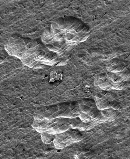
Scanning electron micrograph of ivory surface shows resorption pits where an osteoclast has resorbed the mineralized matrix. Micrograph taken by Western University research technician Tom Chrones at the Biotron Experimental Climate Change Research Centre at Western.
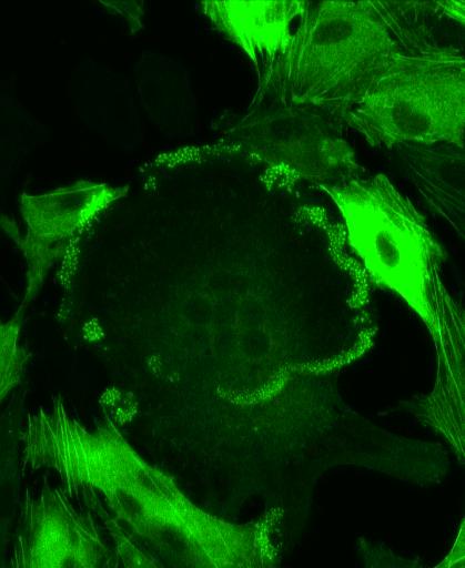
Confocal microscope image reveals distinct patterns of the actin in bone cells. Actin is a cytoskeletal protein that is important for cells to adhere to surfaces, and to move. Actin filaments are stained green. Osteoclast (large multinucleated cell in centre) exhibits prominent actin localization in clusters of 'podosomes' (green puncta) around the edge. This actin network allows osteoclasts to attach to, and cause destruction of the underlying bone. In contrast, conventional actin stress fibers are apparent in neighboring stromal cells. Micrograph taken by Ryan Shugg, first author of the report in the The Journal of Biological Chemistry.


