Zeiss LSM 510-Meta
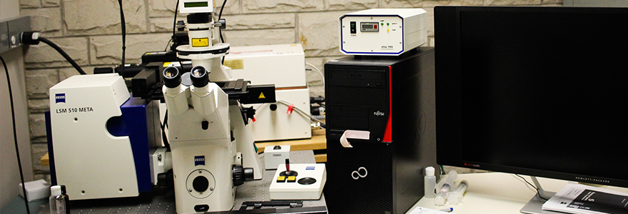
The Zeiss LSM 510-Meta enables users to localize specific biomolecules, analyse FRAP, quantify relative signal intensities, make time and z-series, and do 3-4D life cell imaging microscopy. Furthermore, the META channels enable fluorescent emission fingerprinting to detect up to 10 fluorophores simultaneously.
Key Features
- Seven laser lines: UV 405nm at 25mW, tunable Argon 458/477/488/514nm at 30mW, HeNe1 543nm at 1.0mW, and HeNe2 633nm at 5.0mW.
- Five objective lenses: C-Apochromat 10x/0.45 W, Plan-Neofluar 25x/0.8 Imm corr DIC, C-Apochromat 40x/1.2 W corr, Plan-Neofluar 40x/1.3 Oil DIC, Plan-Apochromat 63x/1.4 Oil DIC.
- One META detector
- Z sectioning (Z-stacking) and 3D reconstruction
Gallery
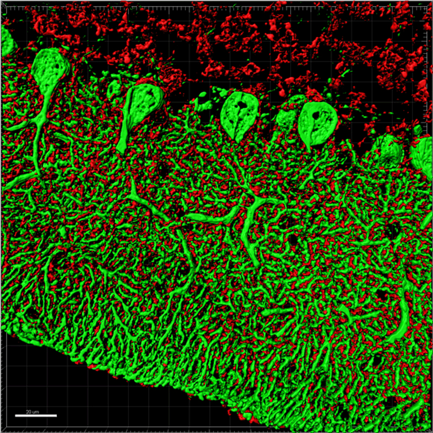
Mouse cerebellum stained with Calbindin (green) amd Synaptophysin (red). This image was captured as a Z-series by Dr. Yanna Xiang using our Zeiss Confocor microscope and pos-processed using Imaris software.
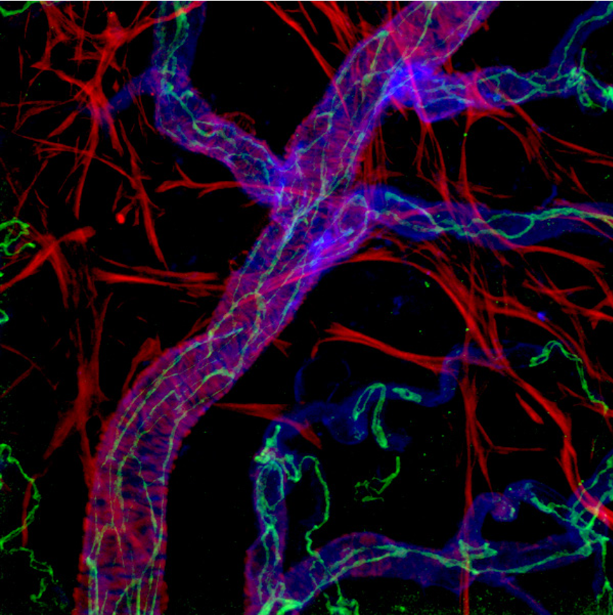
Mouse retinal artery: smooth muscle cells are shown in red; VE-cadherin (marking the boundaries between individual endothelial cells) is shown in green and blue represents isolectin B4 (marking the surface of endothelial cells). This image was acquired by Dr. Hao Yin on our Zeiss Meta 510 microscope.
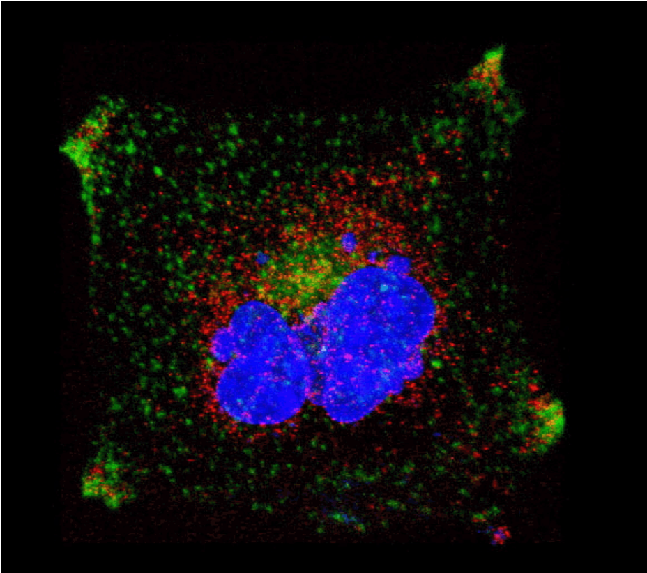
Representative Confocal Image of Cos7 cells labeled for LAMP1-CFP (green), Cox8-dsRed (red) and TOPRO3 (blue). The image was acquired by Dr. Stephen Pasternak on our Meta 510 Microscope.


