EVOS FL Auto 2 Imaging System
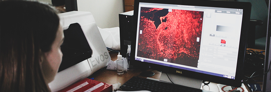
The EVOS fluorescence microscope provides users with exceptional signal-to-noise ratios with its groundbreaking LED illumination system. This system provides top-of-the-line imaging capabilities for cell-based immaging, such as live-cell analysis, image tiling, and Z-stacking. Furthermore, contrary to traditonal mercury-based light sources, LEDs do not require special disposal and are thus more environmentally friendly and energy-efficient.
Automated fluorescence imaging
Key Features
- Four user-changeable fluorescence LED light cubes: RPF, DAPI, CY5, and GFP.
- 10x, 20x, and 40x LPlanFL objectives
- Two cameras: a high-sensitivity monochrome camera optimized for fluorescence imaging and quantitation, and a high-resolution color camera optimized for colorimetric imaging
- Multichannel time-lapse live-cell imaging
- Epifluorescence image capabilities
- EVOSTM FL Auto 2 Software
Gallery
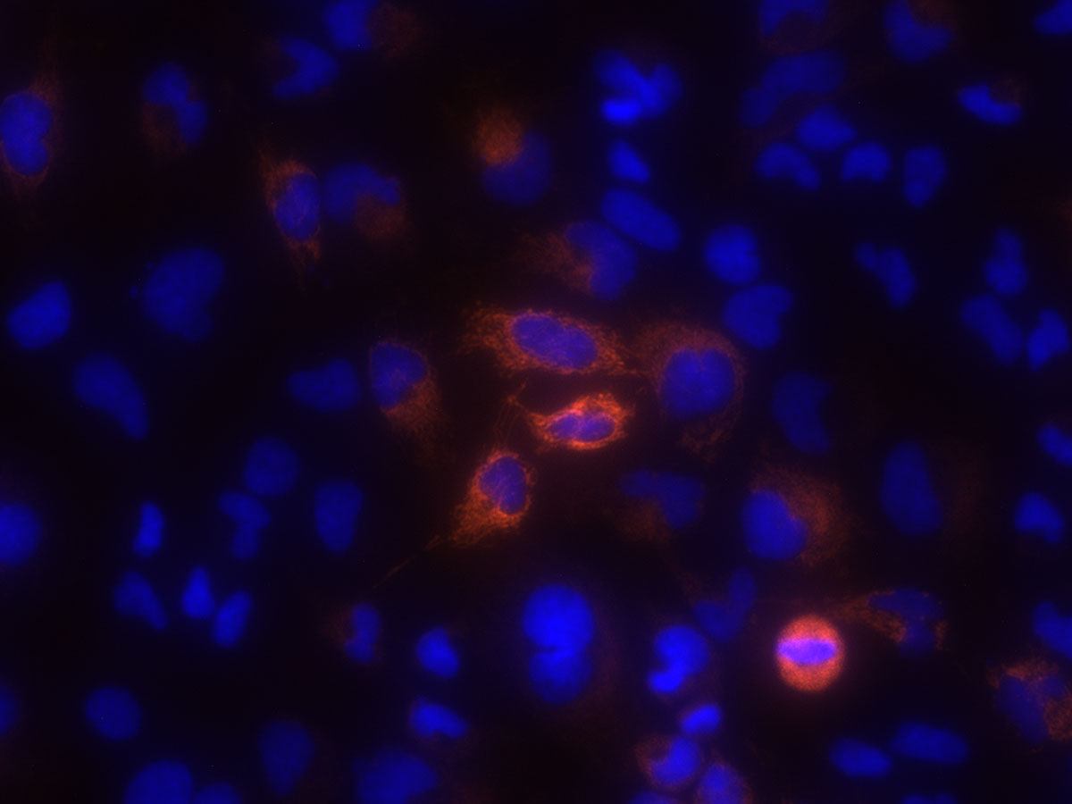
HeLa cells expressing Mitochondria RFP were plated on a 6-well Falcon plate. Cells were then stained with NucBlue Live and imaged in Live Cell Imaging Solution using 40x objective (Thermo Fisher Scientific).


