Gallery
The LRMF is dedicated to providing affordable, high quality, accessible, and user-friendly microscopy services for users in Southwestern Ontario. Below are some of our favourite images captured by users of our facility.
In vivo microscopy scan of a venule and capillaries in the mouse skeletal muscle with plasma tracer (green; FITC-dextran) and leukocytes (yellow; stained with Rhodamine 6G) interacting with venular endothelium. Scan collected with Nikon A1R MP+ Multiphoton Microscope by Stephanie Milkovich, Paulina Kowalewska and Chad Steele.
In vivo 3D scan of a mouse skeletal muscle venule after intravenous injection of a plasma tracer (green; FITC-dextran) and leukocyte stain (red; rhodamine 6G). Captured on Nikon Ti2-E Confocal Inverted Microscope by Stephanie Milkovich and Paulina Kowalewska.
In vivo microscopy of the mouse skeletal muscle microvasculature. Fluorescence 3D scan was acquired after intravenous injection of a plasma tracer (FITC-dextran). Captured on Nikon Ti2-E Confocal Inverted Microscope by Stephanie Milkovich and Paulina Kowalewska.
In vivo microscopy of the mouse skeletal muscle microvasculature. Transillumination technique: light transmitted through the muscle was captured at two wavelength (O2-dependent, top; O2-independent, bottom) allowing for calculation of RBC O2 saturation based on spectral properties of oxygenated and deoxygenated hemoglobin. Captured on Nikon Ti2-E Confocal Inverted Microscope by Stephanie Milkovich and Paulina Kowalewska.
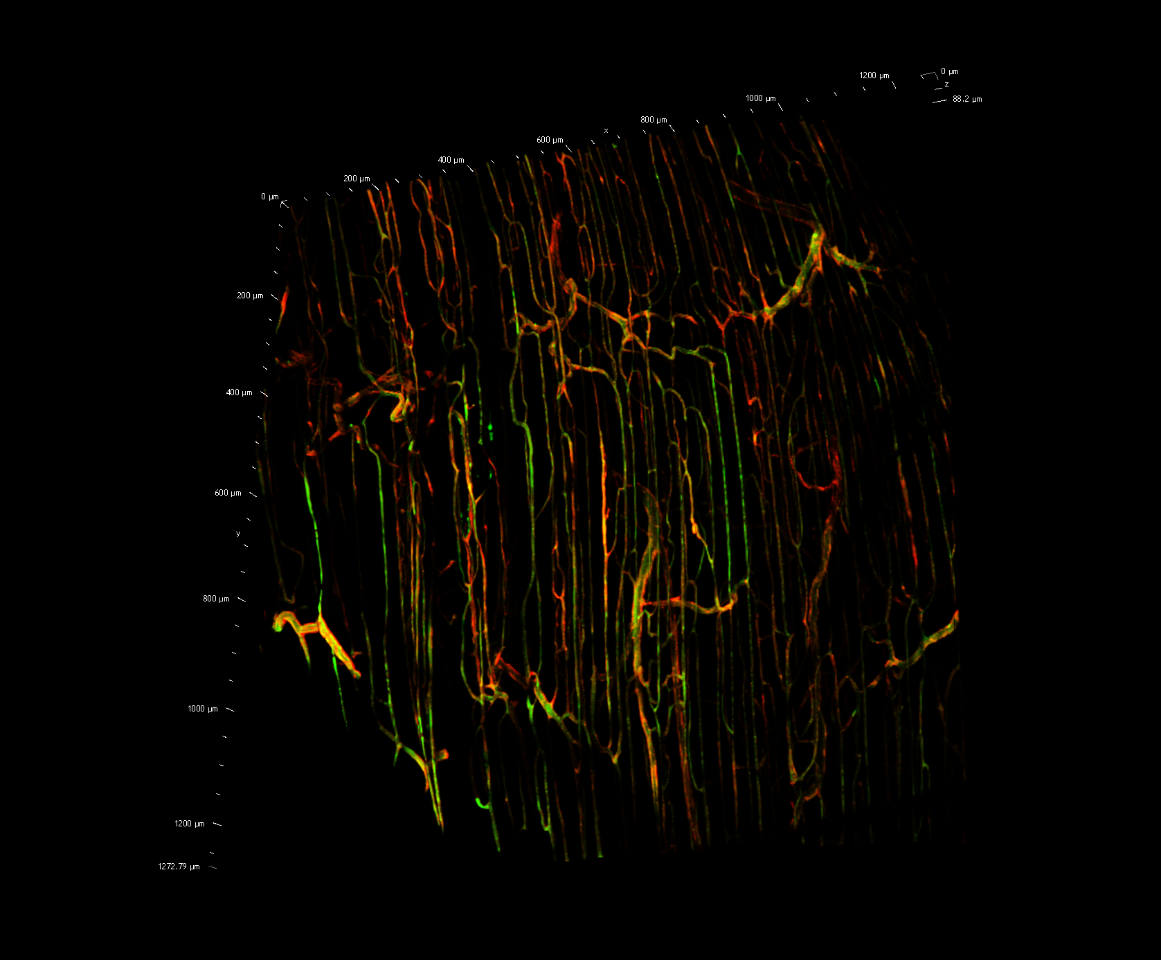
Stitched in vivo image of the mouse skeletal muscle microvasculature with FITC-dextran plasma tracer (green) and CD31-labeled endothelium (red). Captured on Nikon Ti2-E Confocal Inverted Microscope by Stephanie Milkovich and Paulina Kowalewska.
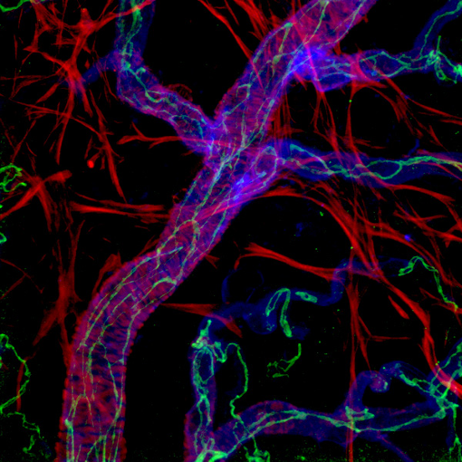
Mouse retinal artery: smooth muscle cells are shown in red; VE-cadherin (marking the boundaries between individual endothelial cells) is shown in green and blue represents isolectin B4 (marking the surface of endothelial cells). This image was acquired on the Zeiss Meta 510 microscope. Image by Dr. Hao Yin.
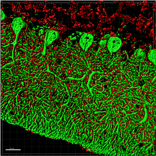
Mouse cerebellum stained with Calbindin (green) and Synaptophysin (red). This image was captured as a Z-series using our Zeiss Confocal microscope and pos-processed using Imaris software. Image captured by Dr. Yanna Xiang.
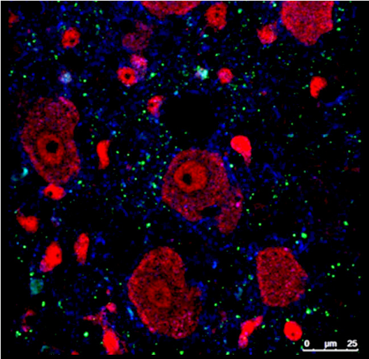
Representative confocal image of brain tissue stained for VGLUT1 (green), BK channels (blue) and giant neurons in the brainstem labelled with NeuN (red). The image was acquired on the Leica SP5 confocal microscope, using 63X objective. Image by Mahabba Smoka.


