

Imaging in motion
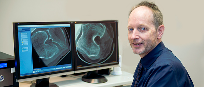
David Holdsworth’s eyes light up when he talks about what the future has in store for studying bones and joints. Holdsworth, PhD, and a team at Robarts Research Institute and Schulich Medicine & Dentistry are working on a project that would allow them to see the human skeleton and all of its intricately connected parts, while in motion.
“Trying to understand the skeleton based on static images of a person lying on a table is like trying to understand consciousness by studying the mind of someone who is asleep,” said Holdsworth.
A recent investment by the Canada Foundation for Innovation is allowing Holdsworth and his colleagues to build a state-of-the art facility at Western University that uses a virtual-reality treadmill to simulate everyday activities, coupled with X-ray technology that can snap up to 30 frames per second.
The result will be the first imaging facility of its kind in the world with the capability of showing exactly how the bones and joints are moving in relation to each other almost in real-time. It opens up possibilities for treatment and diagnosis that Holdsworth says could be game-changing for orthopaedic surgeons.
“Your skeleton is typically in motion, from your spine to your hips to your knees, so we can learn so much more about what’s going wrong if we can get these high-resolution images while the bones are moving,” he said.
It is this certainty that bones and joints need to be studied in their constantly changing state that drives the bulk of Holdsworth’s research program at Robarts. This includes research looking at the complexity of the entire human skeleton right down to how motion affects bone at the cellular level.
Because the skeleton is constantly remodeling itself, Holdsworth and a team of collaborators are also interested in seeing the effect physical stimulation-like exercise has on a bone cell’s ability to regenerate.
Holdsworth and his team are working on building devices that will apply mechanical forces to bone cells while they are being observed under state-of-the-art microscopy. By doing this, they will be able to study exactly what is happening to the cell as it is being exposed to simulated exercise.
They hope it provides clues to developing treatments for conditions like osteoporosis, in which patients experience excessive bone loss.
Although his passion for bones and joints is palpable, Holdsworth wasn’t always interested in this area of work. Schooled in physics and astronomy, Holdsworth changed his focus to medical biophysics based on a desire to improve human health. It was the late Dr. Sandy Kirkley who persuaded him to study the often life-altering conditions of the musculoskeletal system.
“She helped me realize that orthopaedic conditions aren’t just what you get when you’re 72 and you’re retired; these were things that could be life-changing at 23,” said Holdsworth. “One blown knee could mean a whole new career path.”
Tragically, Dr. Kirkley was killed in a plane crash in 2002. She left behind a robust research program that was on the verge of ground-breaking discoveries. Committed to carrying on her legacy, her colleagues were determined to finish the work that she started.
In 2007, the Dr. Sandy Kirkley Chair in Musculoskeletal Research was established, a position that Holdsworth now gratefully occupies.
“She never endorsed the idea of boundaries in research,” Holdsworth said. “That’s how an astrophysicist like me ended up looking at the human skeleton.”


