Search Website
Western’s CFMM marks 30 years of imaging discovery
By Emily Leighton
Researchers have transformed understanding of the brain, healthy aging and disease
By Emily Leighton
Thirty years ago, scientists were only beginning to explore the potential of ultra-high field magnetic resonance imaging (MRI). For Ravi Menon, PhD, the technology offered a way to pursue questions that had previously been out of reach.
“When I started, the motivation was curiosity,” said Menon, professor at Western University's Schulich School of Medicine & Dentistry. “Curiosity to see whether these kinds of scanners could reveal secrets in the brain that we couldn’t see with other tools.”
That pursuit helped establish what would become one of the world’s leading imaging research centres.
For three decades, scientists at Western’s Centre for Functional and Metabolic Mapping (CFMM) have pushed the boundaries of imaging science, helping transform increasingly powerful MRI scanners into essential tools for studying health and disease.
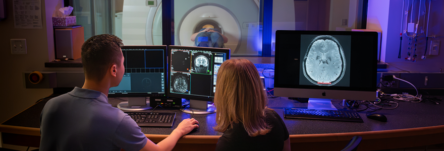
Opened at Robarts Research Institute in 1996, CFMM was uniquely located on the doorstep of Western’s campus and London Health Sciences Centre’s University Hospital – bridging academic and patient care.
“Being next to the hospital was a key feature from the start,” said Menon, CFMM’s founding director. “It created an environment where clinicians and scientists could work closely together to benefit patients.”
When CFMM’s first 4 Tesla scanner arrived that year, it was among the most powerful MRI systems in the world. Today, CFMM houses one of the most advanced suites of imaging technology, including 3 Tesla and 7 Tesla human MRI systems, alongside 9.4 Tesla and 15.2 Tesla preclinical MRI systems for studying animal models of disease.
“We're one of the few labs in the world where all this imaging equipment is one place,” said Menon. “That integration is invaluable. Researchers can move discoveries from animal models to humans – and back again.”
“We’re able to answer questions that many labs would not be able to,” he added.
This range has allowed researchers to study the structure and function of the brain and other organs in unprecedented detail. Ongoing studies are mapping how the brain develops in childhood, how it changes with healthy aging and what goes wrong in neurodegenerative and mental health disorders.
Researchers have advanced understanding of epilepsy, concussion, post-traumatic stress disorder and diseases like Alzheimer’s and Parkinson’s. Their work has also provided a window into what is happening inside the minds of patients in a vegetative state.
"MRI has been revolutionary because it's non-invasive," Menon said. "This ability to image at very high resolution has allowed us to learn things that simply weren't accessibly before – things I never would have imagined possible 30 years ago.
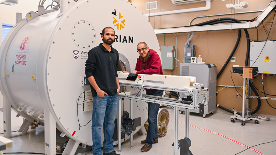
Renil Mathew, a PhD candidate in neuroscience, and Ravi Menon, PhD, in front of the 9.4 Tesla MRI. (Megan Morris/Schulich Medicine & Dentistry)
CFMM’s impact also extends to the people who work and train within its walls. Nearly 1,000 faculty members, visiting scientists and trainees have conducted research through the facility, with former students now leading imaging programs and labs around the world.
As CFMM marks its 30th anniversary, researchers are focused on next-generation high-field imaging, AI integration and the direct measurement of brain activity, with ambitions to expand its role as a national, open-access platform for brain research.
That future is also being shaped by new investments, including the recently announced $8-million endowed Frank and Janice Lochan Neuroimaging Chair for Brain Health to advance the study of neurological disease.
“We need to find what connects all these neurological disorders,” said donor Frank Lochan. “We think neuroimaging is the key to revealing which investigative avenues to pursue.”
For Menon, the questions driving the work remain far from answered.
"There’s so much more to understand,” he said. “We’re going to keep pushing this technology forward, continuing to benefit patients and continuing to unlock the secrets of the brain.”