

Search Website
New CFI funding advances organoid, respiratory imaging research
By Colleen MacDonald
Funded projects include Western's first ever 4D imaging system to advance organoid research
By Colleen MacDonald
Researchers at Western's Schulich School of Medicine & Dentistry have received funding from the Canada Foundation for Innovation (CFI) to advance technologies for health research.
The funding comes through the John R. Evans Leaders Fund (JELF), a core CFI stream that provides up to 40 per cent of the cost of research infrastrucutre, helping universities recruit and retain top researchers.
Among the projects receiving funding is a 4D imaging system for organoid research – the only one one of its kind in Southwestern Ontario.
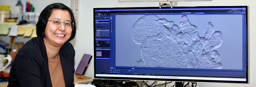
Van Lu, PhD, assistant professor in physiology and pharmacology, received more than $322,000 for the new system. The high-tech microscope and camera system, to be housed at Western, can image complex biological systems in extraordinary detail, offering researchers unprecedented insight into how tissues develop and disease unfolds.
Lu worked with fellow professors Dean Betts, PhD, and Dr. Julio Martinez-Trujillo to bring the technology to Western and make it available to researchers across disciplines. The 4D imaging system is expected to accelerate discoveries in cancer, reproductive health, bowel disease, neuroscience and precision medicine.
“We wanted to take the lead on bringing this equipment to Western, so a wide range of organoid researchers can benefit,” Lu said.
The second JELF-funded project at Schulich Medicine & Dentistry is focused on health outcomes for patients with asthma and chronic obstructive pulmonary disease (COPD). Led by Grace Parraga, PhD, the new funding will support infrastructure upgrades and sustain validated software pipelines, measurements and markers used in respiratory imaghing research.
Parraga's research aims to develop new therapies, reduce health-care costs and train the next generation of respiratory imaging experts.
Advanced 4D imaging technology supports organoid research across Western
Lu, Betts and Martinez-Trujillo collaborate in a research group that studies simplified versions of human organs, known as organoids. These miniature organs, grown in a lab based on human tissues, measure about one millimetre in diameter at most. Though small, the 3D models form all the basic components an organ needs to start developing outside of a living organism.
Using organoids instead of animal models in preclinical studies improves the likelihood of successful treatments that can be moved into clinical trials.
“It’s precision medicine. We can generate many different organoids based on a sample of tissue, test a range of therapeutics or drugs and determine which treatment a person responds to best,” Lu said.
Lu’s organoid research is revealing more about conditions such as colon cancer and inflammatory bowel disease by focusing on gastrointestinal tract development and how it senses nutrients. Betts aims to improve in vitro fertilization outcomes by examining how human stem cell-derived embryo models develop. Martinez-Trujillo is studying how brain stem cells form the earliest stages of neural connections in organoids to better understand neurodevelopmental diseases.
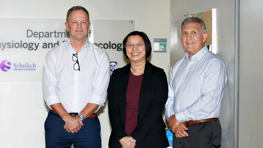
(L to R) Dean Betts, PhD, Van Lu, PhD, and Dr. Julio Martinez-Trujillo put their efforts together to successfully apply for Canada Foundation for Innovation funding that will bring a high-tech 4D imaging system to Western. (Colleen MacDonald/Western News)
The new 4D imaging system will allow researchers to simultaneously examine hundreds of organoids as they develop over the course of weeks or months, instead of analyzing only three-dimensional static images. The system will significantly improve the team’s ability to accurately model living systems by revealing how cells respond to their environment, how they communicate and how diseases begin and progress.
“The advantage is its capacity to capture 3D images over a long period of time, which is necessary because organoids take several weeks to develop,” Lu said. “We can see in real time where the cells we’ve labelled have moved, how many more appear and how they change when we apply a certain treatment.”
AI-powered imaging system positions Western as hub for organoid research
The system includes an incubator to keep the cells growing. Robotics are used to image tissue samples around the clock, while artificial intelligence (AI) built into the system tracks cell changes as they occur. The automation of numerous processes eliminates tedious, repetitive tasks humans can’t do, while allowing multiple researchers to use the system at the same time.
The infrastructure will expand Western’s research impact in biotherapeutics, neuroscience and developmental biology.
Though the 4D imaging system will be installed at Schulich Medicine & Dentistry, it will be available to researchers across Western and its affiliated institutes, bringing together diverse organoid research programs and positioning Western as a hub for organoid research in London, Ont.
“It took a lot of research and development to establish organoids. Now, we’re taking the next step – making organoid models more widely available to researchers,” Lu said. “That could spark new therapies, allow them to be tested relatively quickly and deepen our understanding of the fundamental biology behind cell development.”