Search Website
QUICK LINKS:
Signal in the silence: Researchers uncover hidden potential in discarded MRI data
By Emily Leighton
A new technique is transforming how scientists use fMRI to study the brain
By Emily Leighton
In every functional MRI scan, after the whir and pounding begins, there is a brief 10 to 20 seconds of stabilization as the machine’s magnetic field settles into place. For decades, scientists have treated this period as dead time, discarding the data or ‘dummy scans’.
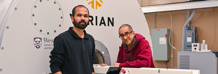
But a team of researchers at Western University’s Centre for Functional and Metabolic Mapping (CFMM) have discovered these early few seconds offer some of the richest data a scanner can produce.
Their new functional MRI (fMRI) technique, published this week in Nature Methods, takes advantage of the brief “start-up” period at the beginning of a scan. By adding short, deliberate pauses – called acquisition-free periods – the researchers allow the scanner’s signal to reset and strengthen, producing sharper, more responsive images of brain activity.

This technique is like adding a turbocharger to your engine. Normally, you throw away the exhaust – here, we’re harnessing that extra energy.
Professor
“This technique is like adding a turbocharger to your engine,” explained Ravi Menon, PhD, the study’s lead author and professor at Schulich Medicine & Dentistry. "Normally, you throw away the exhaust – here, we’re harnessing that extra energy.”
Functional MRI works by tracking tiny changes in blood flow in the brain, revealing which areas of the brain are more active when we think, feel or move. It’s one of the most widely used tools in neuroscience.
The discovery could reshape how researchers study the brain in real time – from mapping memory and attention to tracking seizures or exploring consciousness.
A simple insight, a powerful shift
The breakthrough began as an act of scientific persistence, said Renil Mathew, a PhD candidate in neuroscience and first author on the study.
His PhD project was focused on combining fMRI and electrophysiological data to capture the brain's electrical and blood-flow signals at the same time. The two methods rarely coexist cleanly, so he introduced gaps between scans to record clean electrical data – and noticed something surprising.
“The images right after those pauses were clearer and stronger than usual,” he explained.
By deliberately pausing the scanner to create these windows, then analyzing the high-sensitivity data that follows, the team turned a long-standing limitation into an opportunity.
The approach not only improves data quality, but also boosts efficiency – researchers can achieve the same statistical results with half as many trials.
They also proved the method works across species and field strengths, from 9.4T animal imaging to 3T clinical scanners.
“The physics are pretty simple,” said Menon. “We’re not changing the hardware, just taking advantage of a signal that’s already there.”
Now, the researchers are applying the technique to epilepsy, which could help clinicians better localize seizure activity.
A milestone moment
For Mathew, who began his academic journey studying physics in India before pivoting to neuroscience in Norway, the discovery marks a personal milestone: his first-ever first-author paper – in one of the world’s most prestigious scientific journals.
“I’m glad it happened with my first PhD project,” he said with a smile. “I didn’t expect it to have this kind of impact.”
The work was made possible by Western’s world-class imaging infrastructure – among the most advanced in North America. Within a few steps at CFMM, researchers can move from a 3T human scanner to a 9.4T or even a 15.2T preclinical system. That proximity made it possible for the team to demonstrate the technique’s versatility across platforms and applications.
“In most places, you’d have to go across the country to do that,” Menon said. “Here, it’s literally just around the corner.”
As CFMM approaches its 30th anniversary in 2026, discoveries like this highlight its long-standing role as a hub for curiosity, imaging innovation and cutting-edge technology.
And the findings of this latest study may soon extend far beyond its walls. With only a simple software update, MRI facilities worldwide could adopt the method – transforming moments once considered scientific waste into some of the most valuable data in brain research.
The research team includes Amr Eed, PhD, Martyn Klassen, PhD, Omer Oran, PhD, and Stefan Everling, PhD. The study was funded by the Canadian Institutes of Health Research and BrainsCAN.