

Liver MRI
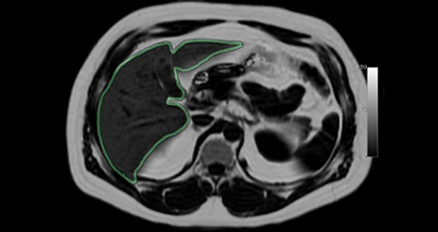
Magnetic Resonance Imaging (MRI) is one of the most diverse imaging modalities in medicine and offers the ability to take images of specific biological tissues. The two most common tissues within the human body are water and fat. Recently it has been discovered that when some organs (specifically the liver) are un healthy, they will contain high levels of fat. In order to diagnose diseases, it is critical to determine an accurate estimate of the amount of fat within the liver (outlined in green).
The current gold standard is to use a liver-biopsy, however this is an invasive method that only samples a small portion of the liver and can lead to misdiagnosis. The objective of our research is to use MRI to quantify the amount of fat within the entire liver. MRI techniques have been developed in the past that are able to form “purely-fat” and “purely-water”(normal liver tissue) images, however this makes the MRI scan longer.
New MRI techniques such as Parallel MRI, allow us to generate a complete image using processing techniques and only a fraction of the data usually required. Using Parallel MRI can speed up an MRI acquisition to less than a “breath-hold”, however it can cause lower quality images if the fraction of data we acquire is not selected carefully. Ultimately, we will compare our MRI fat maps with liver biopsies to determine the accuracy of our non-invasive technique.The increase in scan time is a problem for any type of abdominal imaging, as a patient can only hold their breath for a relatively short time. Therefore in order to produce the clearest image possible, an MRI abdominal scan is limited to the length of a patient “breath-hold”, which is on the order of 20 seconds.
Breakthroughs in MRI are now making it possible to not only image human anatomy, but to also look at human physiology by following metabolic processes. We hope to develop MRI based tools that can track metabolic processes and lead to earlier and more accurate diagnosis of disease.


