

Fetal MRI
Intrauterine growth restriction (IUGR) is a condition that severely compromises the growth of a fetus, resulting in low birth weight fetuses and approximately 1300 fetal deaths per year in Canada [1-3]. Babies born with IUGR suffer from life-long disorders, including neurological deficits, diabetes, and cardiovascular disease [3,4]. Detecting changes in fetal and placental oxygenation, fetal fat distribution and overall fetal volume before birth may help physicians evaluate the risk of growth restriction, stillbirth, and future metabolic health and determine the best time for early intervention, such as induction of preterm delivery. Magnetic Resonance Imaging (MRI) complements routine ultrasound methodologies, by providing a volumetric measurement of fetal adipose tissue distribution.
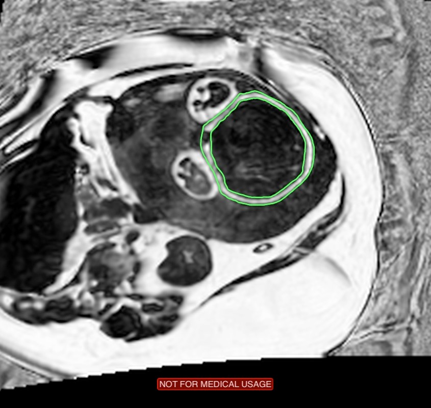
The goal of this project is to develop MRI methods to assess fetal metabolic status. State-of-the art MRI technologies will be employed, including BOLD MRI [5] to measure fetal and placental oxygenation, and water-fat MRI [6,7] to assess fetal adipose tissue distribution. Fetal subcutaneous fat is outlined in green on a fat signal fraction (fat/(water+fat)) image.
 Since MRI is sensitive to motion, methods will be developed to correct for both maternal and fetal motion. Imaging during a breath hold or using respiratory gating will remove maternal respiratory motion, while rapid acquisition methods will be developed to reduce the effects of fetal motion. These rapid methods will be based on combined parallel MRI and compressive sampling, and will aim to obtain images between fetal movements.
References:
1. Statistics Canada HSD. Births 2009. Ottawa, ON: Statistics Canada; 2012.
2. Marsal K. Intrauterine growth restriction. Current opinion in obstetrics & gynecology. 2002; 14(2):127-135.
3. Wu G, Bazer FW, Cudd TA, Meininger CJ, Spencer TE. Maternal nutrition and fetal development. The Journal of nutrition. 2004;134(9):2169-2172.


