The Foster Lab:
Imaging the Invisible

Paula Foster, PhD, is using cutting-edge imaging technology to answer elusive questions about the relationship between immune cells and disease.

By Max Martin, MMJC’19

Paula Foster makes the invisible visible.

Using some of the world’s most cutting-edge imaging technology, she’s discovering new ways for researchers to better track and understand the paths cells take in the body and how that impacts disease.

The imaging specialist has been at the forefront of developing novel technologies and practices for decades, and her lab was the first in the world to show that an MRI could detect single iron-labelled cells.

Now, as part of Western’s Imaging Pathogens for Knowledge Translation (ImPaKT) Facility at Schulich Medicine & Dentistry, she’s heading the first lab in Canada using a brand-new Magnetic Particle Imaging (MPI) machine for cellular and molecular imaging – one of only five in the world.

Imaging expert Paula Foster, PhD, explains how the multi-use molecular imaging equipment inside the ImPaKT Facility will allow for new advances in infectious disease research.

“It’s a brand new kind of imaging,” said Foster, PhD, a Professor in Medical Biophysics and Scientist at Robarts Research Institute. “We have a unique opportunity to lead in this area, we’ve been doing MRI-based iron cell-tracking for so many years, and this will allow us to really improve that.”

paula foster

“What is important but missing right now is the ability to quantify the cells, and that’s where the new equipment comes in. Once we understand the number of cells that are migrating and surviving, therapies can be adjusted, and improvements can be made.”

- Paula Foster, PhD

The MPI machine tracks iron oxide nanoparticles. Various cells, including cancer cells, immune cells and stem cells, are tagged with iron particles to be imaged and visualized in the machine. The image it produces is then overlaid with other imaging scans – like MRI or CTs or X-rays – to get a complete picture of where the cells are in an animal model.

While current MRI technology can image iron, it isn’t able to do so with high specificity and isn’t quantitative – benefits added by the MPI machine, which is housed at Robarts Research Institute. Since the new technology images iron particles and nothing else, researchers will be able to measure the number of cells more precisely than using other methods.

Foster says providing a cell number is imperative for cell therapy research.

“What is important but missing right now is the ability to quantify the cells, and that’s where the new equipment comes in,” she explained. “Once we understand the number of cells that are migrating and surviving, therapies can be adjusted, and improvements can be made.”


ImPaKT equipment mpi
A dual MRI/PET scanner is one of two multi-use imaging machines in the Facility.
The MRI provides an anatomical reference, while the PET scan shows important molecular information.
Did you know?

In Canada:
• Cancer is the #1 cause of death
• Every hour, 25 people are diagnosed with cancer
• 1 in 2 people will develop cancer in their lifetime
• 1 in 4 people will die from cancer

*Statistics from the Canadian Cancer Society

But the MPI machine is just one tool in an arsenal of many at the disposal of researchers in ImPaKT.

ImPaKT is home to one room that features two multi-use imaging machines that will allow researchers to conduct up to four different imaging scans.

“It’s pretty unheard of to have four different types in one small area together,” Foster said. “It’s pretty much all you could want, and each gives you different information.”

The first instrument is a dual MRI/PET scanner. MRI provides anatomical information, while the PET scan offers molecular information. The second machine does bioluminescence imaging, which tells researchers about cell viability. Its dual function is as a CT scanner, which provides an image of bones, blood vessels and soft tissue.

Combining all this information along with newfound insight from the MPI machine could offer a deeper understanding of how various cells in the body respond to pathogens or disease.

“Depending on what you’re studying, you can use all four modalities one after the other in the same room to collect a lot of information,” Foster said. “The goal is to be imaging cells that are involved in disease much earlier.”

ImPaKT equipment mpi
Paula Foster, PhD, and John Ronald, PhD, work inside the imaging suite in the ImPaKT Facility.
The Level 2+/3 containment room houses four different imaging modalities.
“These will be experiments that we’ve always wanted to do, but we’ve never been able to do because of the barriers of working with viruses and pathogens outside of a containment facility.”
– Paula Foster, PhD

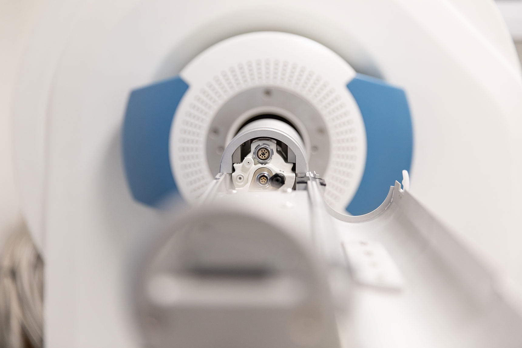
Another benefit is that the equipment is housed inside ImPaKT’s Level 2+/3 containment facility, meaning live, infected animal models can be imaged.

“It’s completely unique in Canada, and it’s very rare. Nowhere have people been willing to put this huge investment in imaging equipment into a facility like this,” Foster explained.

The imaging suite’s containment level is adjustable, meaning the room can transition from level 2+ to level 3, depending on the experiments being conducted and the pathogens being worked with.

“These will be experiments that we’ve always wanted to do, but we’ve never been able to do because of the barriers of working with viruses and pathogens outside of a containment facility.”

Did you know?

In Canada in 2019:
• An estimated 220,400 people were diagnosed with cancer
• An estimated 82,100 people died of cancer

*Statistics from the Canadian Cancer Society

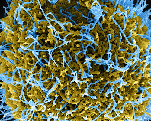
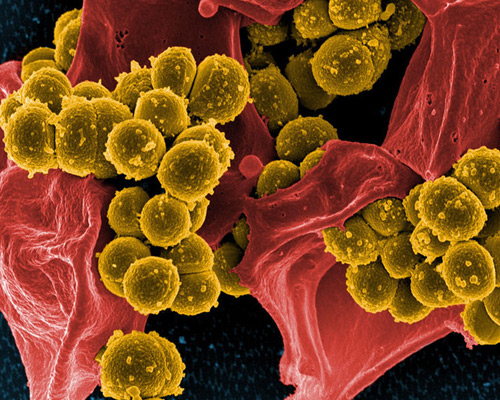
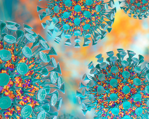
Since the MPI technology is so new, Foster is working on ways to develop and enhance its capabilities. This will allow for improved study into the area of cell therapy, such as stem-cell-based regenerative medicine for treating arthritis or multiple sclerosis. MPI technology also has the potential to help develop novel therapies for diseases like HIV/AIDS, Zika virus and staph A.

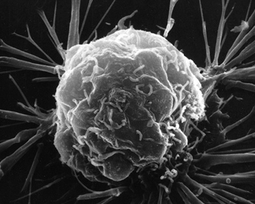
One application Foster is currently studying with the MPI technology is cancer immunotherapy.

In immunotherapy, a patient’s immune cells are removed and modified to better fight the patient’s cancer before being reinjected back into the patient. From the injection site, the cells must travel to the lymph nodes to interact with other cells that do the cancer-killing.

“There are a lot of questions that remain about cancer immunotherapy, and a lot of them are about how many cells we should be injecting, where to inject them, and how long it takes for them to migrate to the lymph nodes,” Foster explained.

This is where imaging iron-tagged cells with MPI will be essential to track the number of cancer-fighting immune cells that make it from the injection site to the lymph nodes. The results from these pre-clinical studies can be sent to clinicians so they can modify their protocols.

“It can directly impact the patient’s outcome because the number of cells that get to the lymph nodes is directly related to the patient’s response to these types of treatments,” Foster said.

“The whole field of imaging infectious disease is just beginning. This will al be entirely new, every experiment that’s done in ImPaKT will be new, and that’s what makes it so exciting.”
– Paula Foster, PhD
ImPaKT equipment mpi
John Ronald, PhD, and Paula Foster, PhD,
work inside the Facility’s imaging suite using a 3T PET/MRI imaging machine.

As imaging technology continues to develop, Foster expects cell tracking to one day play a critical role in clinical settings. “Imaging will become a really important tool at the bedside for all forms of cell therapy,” she said.

The imaging equipment in the ImPaKT Facility is set to change how researchers and clinicians fight infectious disease and will offer up new clues about the immune response. But the true potential of the Facility is just at its dawn, as researchers embark into new frontiers in science and discovery.

“The whole field of imaging infectious disease is just beginning,” Foster said. “This will all be entirely new, every experiment that’s done in the ImPaKT Facility will be new, and that’s what makes it so exciting.”


“Imaging will become a really important tool at the bedside for all forms of cell therapy.”
– Paula Foster, PhD

Read other stories in this series