

New study expands understanding of brain blood flow and neurological disorders
By Cynthia Fazio
The hippocampus – a seahorse-shaped region of the brain which plays a particularly important role in cognitive aging and memory function – has been studied as a singular region for several years. However, there remains a gap in understanding the factors underlying age- or disease-related changes between the different regions of the hippocampus, or subfields, until now.
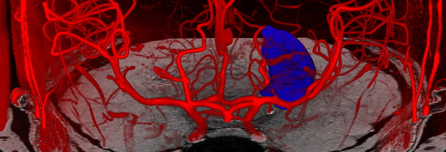
A study by researchers from Western and Maastricht University in the Netherlands published today in the high impact journal PNAS, provides new insight into what perfusion, the flow of blood to tissues, looks like in a healthy brain. The study explains how the subsequent hippocampal perfusion map, a map displaying the distribution and amount of blood in the tissue, can serve as a baseline to compare with a hippocampus that has signs of damage.
The team of researchers developed an efficient, non-invasive method to better understand perfusion in various regions of the hippocampus which could aid in the diagnosis of neurological diseases such as Alzheimer’s, epilepsy and schizophrenia, as well as help promote healthy aging.
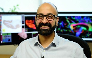
 Top: Ali Khan, PhD, associate professor in the department of Medical Biophysics.
Bottom: Roy Haast, BrainsCAN postdoctoral associate
To get a clearer picture of the structure and function of this crucial region in our brain, the team, involving Ali Khan, PhD, associate professor in the department of Medical Biophysics, and co-led by BrainsCAN postdoctoral associate Roy Haast, developed a process called high-resolution 7 tesla (7T) arterial spin labeling (ASL), a non-invasive magnetic resonance imaging (MRI) method that facilitates measurements of blood flow in the hippocampus.
They also implemented time-of-flight magnetic resonance angiography (TOF-MRA), a non-contrast-enhanced MRI technique to visualize flow within vessels, to see the differences in the size (diameter) and positioning of arteries and to assess their impact on blood flow. Lastly, they used HippUnfold technology – an open-source web-based app, co-developed by Khan, modelling the hippocampus as an unfolded surface – to enable accurate and precise assessments of tissue perfusion in the hippocampus.
“We leverage advanced computational methods, called unfolding, to characterize the perfusion. This has not been done before in a living human,” said Haast. The study involved eleven healthy participants.
MRI scans for the brain typically take 20 minutes to an hour. The 7T MRI ASL protocol implemented by the researchers, by contrast, only takes five minutes, and can capture higher quality images showing patterns of the blood flow to inform patients of the health of their brain. This has the potential to improve ease and accessibility for physicians to check the health of a patient’s hippocampus especially if there is a familial history of neurological diseases and can help with early detection.
There are five hippocampal subfields, of which one, labelled CA1, is typically the region where clinicians find damage, explains Khan, who is also a Canada Research Chair in Computational Neuroimaging and director of the Khan Computational Imaging Lab at Robarts Research Institute.
“Our findings show that even in in healthy adults, there's lower perfusion in CA1. So, it might have a lower threshold for insult when the hippocampus is affected and could point to what might be responsible for why the hippocampus is so ubiquitous in all these different disorders.”
These findings will aid the community in interpreting changes in the hippocampal subfields relevant to neurological diseases and cognitive neuroscience and provide an imaging framework to guide researchers in setting up protocols when studying perfusion in various regions of the hippocampus.
“Our imaging shows precise and accurate measurements of what the normal hippocampus physiology is, providing a basis to compare with abnormal physiology. It is also fast and requires a small sample size,” said Haast.
By quantifying blood flow across hippocampal subfields, it is possible to better understand the normal patterns and how they relate to specific functions. This study provides an MRI imaging framework that allows for characterization of perfusion in a living human and provides a baseline for comparison with diseased states such as Alzheimer’s, dementia, temporal lobe epilepsy and schizophrenia.
For next steps, the researchers hope to use these techniques in more clinical studies, and to characterize the changes in abnormal conditions, which could lead to earlier detection and assessment of the progression of these diseases, better treatment and better health outcomes.


