

Taking a closer look at disease
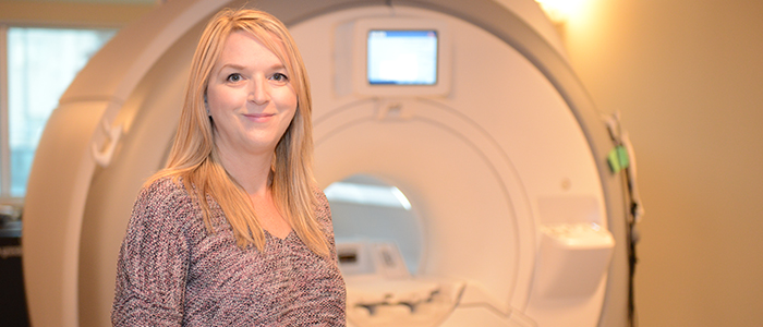
When Paula Foster, PhD, was completing her MSc in Biomedical Sciences at the University of Guelph, she had the opportunity to work on a project with the Ontario Veterinary College that involved magnetic resonance imaging (MRI). Little did she know that imaging the animals that came into the College would prepare her for her current role as the leader of the Cellular and Molecular Imaging program at Robarts Research Institute.
“I think I always knew that I wanted to do research, but I’m certain I didn’t know what type of research at the time,” Foster admitted. “The veterinary college had a big role in generating the interest and enthusiasm for what I do now.”
After completing her two-year postdoctoral fellowship at Robarts, Foster was hired on as a scientist in 2000. She has since been working on a new imaging technique that is referred to as “cellular MRI” or “cell tracking with MRI” — a technique that has not been used anywhere else in Canada.
“In the first five years that I was working here, I was working with other scientists in the imaging lab to developing the technology — the hardware and software — to image really small things with MRI,” she said.
“With MRI, what people think about is imaging a tumour or a lesion or an injury of some type, but what we’re doing is trying to image cells or cellular events that would actually be considered pre-disease or early detection of the cellular events that led up to the tumour or the lesion.”
According to Foster, her team will label the cells they want to track with an agent that is detectable with MRI. Most of the work she has completed has been with iron nanoparticles.
Approximately 40 per cent of what Foster and her team do in the lab involves studying stem cells that might be used to treat several diseases. Tracking stem cells used for tissue repair and regeneration can be applied to multiple sclerosis, spinal cord injury, and peripheral vascular disease.
The remaining 60 per cent of her research involves either tracking the metastasis or movement of cancer cells, or tracking the cells that are used to treat cancer, which is called cancer immunotherapy.
While Foster is happy with the work her lab has accomplished so far, the next big thing they would like to see is this research being moved into clinical trials. According to Foster, MRI is a great imaging tool because it’s non-invasive and it’s not harmful, so patients can be imaged multiple times.
“Cell tracking with MRI in patients has not been done in Canada yet, so we want to be the first to do that for sure, that’s the main goal,” she said. They currently have funding to use MRI to track immunotherapy in prostate cancer patients.
When asked what inspires Foster to continue working in this field, her answer is simple and straightforward: she continues to learn something new every day.
“For me, this particular field of research has so many different applications and possibilities because we’re talking about cells in disease," she said. "There are so many different things I could do with that."


