Behind the door: London Regional Flow Cytometry Facility
We’re taking a look behind the doors of Schulich Medicine & Dentistry’s core facilities. This month, we toured the London Regional Flow Cytometry Facility (LRFCF), based at Robarts Research Institute, with manager Kristin Chadwick, PhD, who explained the technology, equipment and services the LRFCF provides to the London research community.
What exactly is flow cytometry?
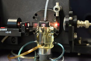
Flow cytometry is a technique for the high-throughput examination of microscopic particles in a fluid stream. It’s a laser-based technology that enables us to analyze cells or particles by detecting fluorescent reagents, called fluorophores, on or within a cell particle.
Fluorophores come in a range of different colours, and can be excited by lasers of different wavelength. We can then identify different cell types based on the various colours they emit.
 Fluorophores can be linked to antibodies that bind to proteins on or within a cell.We may also use fluorescent probes to bind to membranes or DNA, or which change colour in response to physiological stimuli. In other types of experiments,fluorescent proteins (for example, green or red fluorescent proteins - GFP or RFP) are expressed in the cells as markers of transfection or viral infection. Our flow cytometers allow you to look at one, two, or up to 13 colours at one time on a single cell or particle.
Flow cytometers can process up to 20,000 cells per second, which is why we call this high-throughput examination. If you have cells or particles that can be processed down to the single-cell level, then this is a really powerful technology for exploration.
What type of investigations require flow cytometry? What research questions are people who use the facility trying to answer?
We provide access to two different classes of instruments at the LRFCF: analytical flow cytometers and a cell sorting cytometer.
Flow cytometric analyzers allow investigators to conduct end-point analyses, where all of the experimental work is complete and the flow cytometer provides a snapshot of what the cells looked like (their phenotype) or what they were functionally doing at the conclusion of an experiment. Time course analyses are possible by harvesting cells at different time points during an experiment to look at changes in cell phenotype or function.
Our flow cytometric cell sorter instead plays a role at the beginning or mid-point of an experiment. This powerful instrument allows an investigator to physically isolate multiple cell types from a complex population in a viable, sterile manner, for downstream investigation. Cells collected in this manner can be placed back into culture, injected into an experimental animal for further assessment, or can be used as a source of protein or genetic material for further study. We can sort cells as bulk populations into collection tubes, or using the automated cell deposition unit (ACDU) we can place defined numbers of cells onto slides, or into specific wells of multi-well plates.
Our user group is comprised of a wide range of investigators from most departments at Schulich, as well as members of the basic science departments.
- We serve immunologists, who examine, or isolate, various sub-sets of cells active during immune response. For example, during HIV/AIDS disease progression or transplant rejection.
- We serve is stem cell biologists who are examining very rare cell sub-sets of normal and cancerous stem cells (SC), including hematopoietic SCs, Embryonic SCs, Mesemchyml SCs, to name a few.
- We serve physiologists examining changes in cell cycle progression or apoptosis in response to drug treatments or gene knockouts.
- We serve microbiologists who wish to examine the activities of yeast, bacteria, or bacteria-infected cells in a large number of assay systems.
- We serve investigators from a number of departments who need to track cellular transfection efficiency or virus infection through detection of a wide range of fluorescent proteins.
In fact, the fastest growing demand for our cell sorting service specifically, is single-cell cloning for researchers using CRISPR-Cas9 technology, as well as more traditional transfection and viral transduction experiments. Here, plasmids to inactivate, knockout or knockin a particular gene are delivered into a cell population along with a fluorescent reporter gene. Two to three days after gene delivery, these cells can be brought to the LRFCF, where our cell sorter can quickly and efficiently deposit single fluorescent cells into 96-well plates for generation of clonal cultures that can be scanned for a successful genetic manipulation. Each plate can be processed in 5-10 minutes, allowing a number of cell lines to be quickly processed in this manner. This is a much more efficient way to generate purified populations, weeks to months faster than they can generate using the traditional cloning and selection methods.
In short, as long as there is a way to add a colour to a cell as an indicator of function or phenotype, flow cytometers can allow an investigator to examine or isolate those cells for further investigation.
Who can access the LRFCF and what services does it provide?
The LRFCF is open to anyone in need our services or expertise from the greater London and area research community. For the flow cytometer analyzers, we offer a comprehensive new user training program. Anyone can be trained for independent analytical cytometer operation. Graduate students, staff and investigators can receive 24/7 access to the facility’s analytical cytometers (and associated computers with analysis software) upon completion of training, while undergraduate students and volunteers are allowed supervised access to the cytometers and computers. Our training program includes all steps of cytometer operation, including troubleshooting, to ensure our users are comfortable with independently generating their data.
For investigators requiring infrequent or sporadic cytometry analysis, we also offer an operator assisted analysis option, where I will acquire and analyze the data for an additional fee.
Cell sorting is offered by appointment only, and I act as the only dedicated operator of the cell sorter. Anyone who needs cells sorted will prepare their cells in their own lab, bring them to me for isolation. Together we will identify their populations of interest, and then they are free to return to their lab while I process their samples. At the end of their appointment, a user will walk away with a collection of tubes, slides or plates containing their cells of interest.
Last August, biosafety cabinet was installed over the cell sorter. This was an important upgrade that allows us to now safely isolate cells containing a wide range of infectious agents, in addition to providing a greater assurance of sample sterility for all cell types. This cabinet allows us to confidently sort a number of cell types we couldn’t work on before, including live bacteria or virus infected cells, after appropriate biosafety approval.
What other upgrades or changes are coming to the LRFCF?
We have established a partnership with the new Imaging Pathogens for Knowledge Translation (ImPaKT) Facility. The ImPaKT team will be installing an analysis cytometer in their facility, to allow users to analyze infectious samples in the Level 2+ containment area where these samples are being generated. An identical machine will be installed here at the LRFCF, enabling us to train ImPaKT Facility users in a safe environment.
What does your role as the facility manager entail? And who are the other team members?
I am the only staff member here, so I look after the machines, complete quality control and take care of the regular cleaning and service needs. I also manage the training for all of the facility’s users, and look after the administrative duties, such as billing, budgeting and stocking supplies.
Greg Dekaban, PhD, is the LRFCF’s Scientific Director, and David Hess, PhD, is the Co-Director. Greg’s CFI research grant funding allowed the purchase of our high-end LSR II cytometer in 2010, in a critical infrastructure upgrade. Subsequently he was able to secure funds from Schulich and Robarts for the Canto cytometer to further upgrade cytometer infrastructure. David’s stem cell work is entirely dependent on cell sorting, and it was his CFI grant that secured the purchase of our most recent cell sorter, after our last sorter retired due to obsolescence.
Members of both Greg and David's lab arekey users of our facility, so they both know what the facility needs to help everyone meet their experimental goals. And both have offered the time and services of key members of their staff to help keep the facility running smoothly when I am offsite. It’s great to have them as resources.
What’s the most exciting aspect of this work for you?
In my role, I get to see the wide variety of research that labs in the London region are involved with, and it's very rewarding to help them work through their research problems. The work I did during my PhD training was based on flow cytometry and cell sorting, and it’s really exciting to see these diverse projects using technology that was so critical to my own training.
What do you want people to know about the facility?
For those looking to make use of the facility, my advice is to plan ahead and connect with me well in advance. People tend to wait until the day or week they need our services. However, since our instruments are heavily used and my time is in high demand, they may be faced with up to a four-week wait for services at our busiest periods.
The full training for a user to become independent on our analyzers could take weeks to months, depending on how frequently they can generate experimental samples for instruction, and how quickly they are able to optimize their protocols. Since it can take a while before users are proficient, planning ahead is key. And for cell sorting, the larger the sample or longer the sort, the more notice is required to obtain an appropriate appointment time.
Individual labs are now purchasing their own cytometry equipment. What advantages does the LRFCF provide to researchers?
Cytometers are happiest when they are being frequently used and well maintained. As cytometers age, they can become very expensive to maintain in good working order. Without someone on-site dedicated to maintenance, training and oversight, it’s much harder to generate good quality data. We have the dedicated technical expertise to troubleshoot these machines, and our instruments are quality control checked every single day. We rarely experience down time due to instrument malfunction. Due to collection of user fees, we have the funds available to ensure that all instruments are kept in a state of good repair.
As well, in a shared environment such as this, a wide range of users come in with a variety of experiments. This is an atmosphere where both myself, and other proficient users, can be easily found for assistance or advice with experimental execution or troubleshooting.
Furthermore, we are the only core facility in the region that has a cell sorter with a dedicated operator. With more than 12 years of experiences with cell sorting, we can quickly and efficiently isolate a wide range of cell types, from easy to more difficult samples.
Finally, our cytometers are purchased with the goal of facilitating the widest possible range of experiments and a heavy use environment. Our cytometers in general are equipped with more lasers and more detectors than the smaller instruments that are often in individual labs. These instruments allow the use of a wide range of fluorophores. The best example being that we ensure all of our cytometers come equipped with a yellow-green/yellow laser (561 nm) that allow the detection of a wide range of red-shifted fluorescent proteins that you simply cannot see on the smaller, two and three laser cytometers that are being installed in individual labs.


