

A New Picture of Chronic Lung Disease
A novel approach to imaging is leading to changes in the way asthma and COPD are understood and treated
By Pat Morden, BA’77
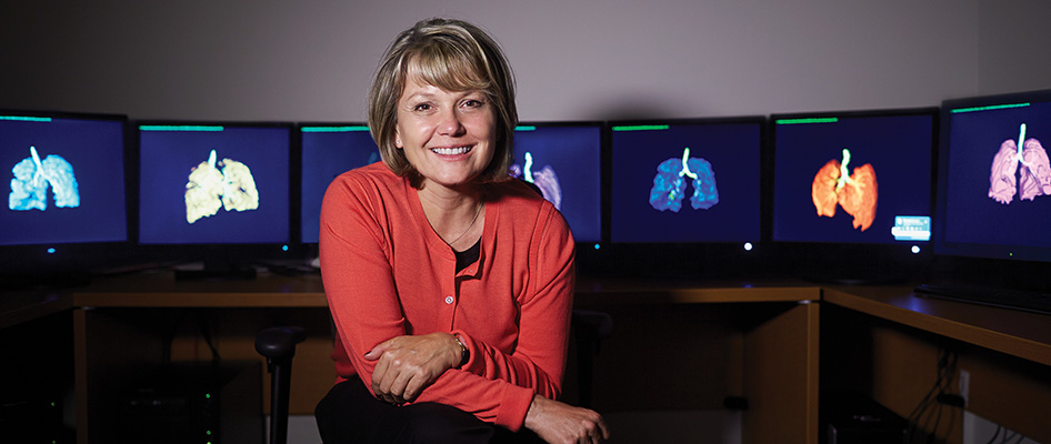
When Grace Parraga, HBSc’84, MSc’86, PhD, talks about Javier Avina, you can hear the frustration in her voice.
Avina is a 10-year-old Wallaceburg boy who died during an asthma attack at school in early April 2016. Parraga believes that the innovative imaging technique developed by her team can be used to predict and perhaps help prevent severe asthma attacks like Avina’s.
Parraga is a scientist who started her career using nuclear magnetic resonance spectroscopy to study protein structure and function, and protein/DNA binding. “I was trained as a hard core basic researcher and determined some fundamental protein structure-function findings,” she said. After completing post-doctoral training in Switzerland, she joined a major multinational pharmaceutical company “in the gap between basic and clinical research.” She joined Robarts in 1998, first as a research administrator and then as a scientist in 2004.
Today she and her team work in a facility created in part through the philanthropy of Richard (LLD’79) and Beryl (LLD’97) Ivey. “They wanted a space where imaging research could be tested in patients and translated into clinical practice,” she said. “This was their dream and vision.”
Under Parraga’s leadership, a remarkable program has taken shape and is making important advances. “Grace is leading the world in lung imaging innovation,” said Dr. Chris Licskai, a respirologist who collaborates with the program. “She has developed a novel tool that has great promise.”
Chronic lung disease is one of the leading causes of death and disability in Canada. More than 2.5-million Canadians, most of them children, live with asthma. It’s also estimated that 1.5-million Canadians have chronic obstructive lung disease (COPD), including one in four hospitalized patients in Ontario.
The standard clinical method for measuring lung function involves quantifying airflow using a spirometer. In the clinic, patients with asthma or COPD blow into the spirometry device and, in some cases, chest X-rays and CT images are also used to help better understand the disease.
Parraga and her team have developed a technique called pulmonary functional Magnetic Resonance (MR) imaging. During testing, the patient breathes in a novel gas contrast agent (produced on-site) while in the MRI scanner. Within a few seconds a high resolution three-dimensional image is produced that shows where the inhaled gas goes and where it can’t go due to airway and other lung abnormalities. “This technique gives us a really good ability to correlate lung structure and function,” said Dr. David McCormack, a respirologist and another clinical collaborator in the program. “That’s pretty unique and a powerful tool.”
The inhaled gases used are not radioactive and are completely inert, so the test is safe and well-tolerated by patients. As a result, it’s possible to image patients before and after taking their medications, before and after walking, and before and after an induced asthma attack.
“If you want to have a leading program, you need a leader with the motivation, skills and imagination to build it. Grace has been able to take a fascinating new imaging technique and build a bridge to the clinical world.” —Dr. Chris Licskai
The images produced by this technique have led to a major shift in the understanding of lung disease. “We used to think about asthma as a fairly homogeneous disease, affecting all areas of the lung to a similar degree,” said Dr. McCormack. “Our data have revealed that it’s a very regionally heterogeneous disease.”
Similarly, COPD affects some parts of the lungs more than others. One analogy, Dr. McCormack says, is to coronary artery disease, in which some, but not all, arteries are blocked with atherosclerotic plaque.
This understanding, in turn, is leading to new therapeutic approaches. “Clinically we’re moving toward a better understanding of the individual phenotype in these diseases,” said Dr. Licskai. “These images may help us to personalize therapy.” For example, the program is part of a study on the use of thermal ablation delivered via a bronchoscope. The goal is to improve asthma control by reducing smooth muscle in the airways. Right now, the whole lung is treated. Knowing where the problem areas are in each patient could lead to a more targeted approach.
The technique is also helping to account for unexplained symptoms reported by patients.
“We have patients who don’t appear to be severely ill, based on spirometry measurements but they have a terrible quality of life,” said Parraga. “When we image them, we can see why.”
The images can also be used to track disease progression, and to identify biomarkers of severe disease that lead to repeated hospitalizations. They can also be used to measure the efficacy of potential new treatments and delivery methods. Ultimately, they may shed light on many other questions about lung disease, including why some people with asthma develop a form of permanent lung damage via remodelling of the airways.
Parraga says there is still some work to do before the technique becomes standard clinical practice worldwide and in Canada. A health economics project is currently under way with collaborators across Canada. “We have to justify, based on patient outcomes, the rationale for MRI in these patients,” she said. “But with our approach, patients are only in the scanner for 10 seconds which will certainly help to contain costs. Improved treatment could lead to fewer hospitalizations,” she added. The average cost of a hospital stay due to COPD is estimated at $10,000.
Parraga’s leadership is a key factor in the program’s remarkable success, said Dr. Licskai. “If you want to have a leading program, you need a leader with the motivation, skills and imagination to build it. Grace has been able to take a fascinating new imaging technique and build a bridge to the clinical world.”
Dr. McCormack agrees, adding, “If you have the technology but not the patients and clinical expertise, it doesn’t work as well. Here we have a really nice interaction between basic scientists and clinicians. And it’s working.”


